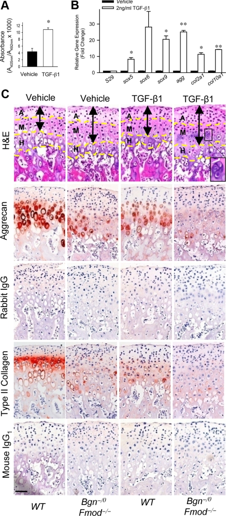
Figure 4.
TGF-β1 accelerates both formation and degeneration of mandibular condylar cartilages in the absence of Bgn and Fmod. A: TGF-β1 induces proliferation of MCCs. BrdU ELISA was used to measure proliferation of MCCs treated with vehicle or TGF-β1. Data are mean ± SEM of 6 wells. *P < 0.005 vehicle versus TGF-β1. B: TGF-β1 induces the expression of chondrogenic-related genes in the MCCs. Quantitative real-time RT-PCR analysis compared the expression levels of chondrogenic-related genes using total RNA isolated from MCCs treated with vehicle or TGF-β1. Gene expression was normalized to the housekeeping gene S29. The expression of genes in MCCs treated with vehicle was relative to that in MCCs treated with TGF-β1. Data are mean ± SEM of 2 experiments. *P < 0.02, **P < 0.002 vehicle versus TGF-β1. C: TGF-β1 accelerates loss of aggrecan content in mandibular condyle explants. Whole mandibles were isolated from wild-type and Bgn−/0 Fmod−/− mice and explants were cultured for 48 hours with vehicle or 2 ng/ml TGF-β1. Comparable histological sections from explants were stained with H&E. Aggrecan and type II collagen expression was examined by immunohistochemistry. Rabbit IgG and mouse IgG1 were used under the same conditions as negative controls, respectively. Dashed yellow lines divide the condyle into articular zone (A), mature zone (M) and hypertrophic zone (H). Black double-ended arrows indicate extent of articular and mature zones. Black square indicates chondrocyte cluster. Scale bar = 50 μm.

