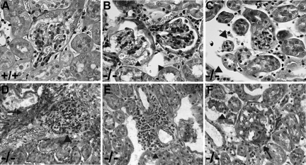
Figure 1.
Light microscopy. Paraffin sections were stained with periodic acid–Schiff. Samples from wild-type (wt) (representative glomerulus shown in A) and lama4−/− (ko) mice (B and C) at 7 weeks of age and from lama4−/− animals at 6 (D), 9 (E), and 18 (F) months of age are shown. Dilated glomerular capillaries (B) and peritubular capillaries (B and C) are shown (arrowheads). Note the proliferative changes in the glomerulus in D (arrowhead), as well as the area of tubulointerstitial fibrosis (arrow). A perivascular inflammatory infiltrate is shown in E (arrowhead). Glomerulosclerosis (arrow) and tubular atrophy (arrowhead) are apparent in F. No glomerular or tubulointerstitial fibrosis was seen in wild-type mice up to 23 months in age.

