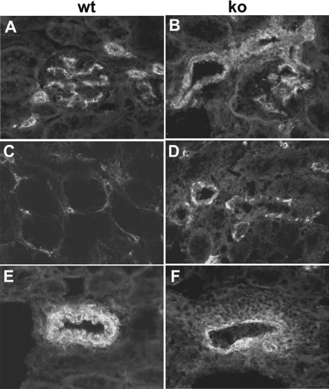
Figure 5.
Pericyte staining. Samples from wt (A, C, E) and ko (B, D, F) mice were stained with antibody to NG2. Note the increased staining in the extra-glomerular mesangium and around afferent and efferent arterioles in B. Note the enlarged vessels with discontinuous pericytes in D. Note the large number of pericytes that surround, but are distant from the vessel wall in F.

