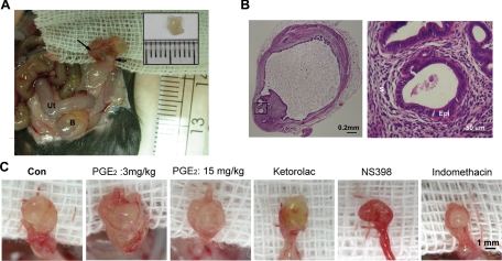
Figure 5.
Macroscopic and microscopic images of endometriotic-like lesions in female C57BL/6NCrj recipients after syngeneic uterine tissue transfer. A: Four weeks after injection, a recipient displayed significant gross vascularity and hyperemia within the lesion and marked vascular recruitment along the periphery of the peritoneal attachment site (arrows). Ut: uterus; B: bladder. The inset shows the endometrial tissue peeled from a donor mouse. Eight pieces of such endometrial fragments were injected into the peritoneal cavity of each recipient. B: Representative images showing H&E–stained adhesive endometriotic tissues in a surgery-induced endometriotic mouse. The right panel is a high-magnification image from the square indicated in the left panel. S: stroma, Epi: epithelium. C: Macroscopic images of endometriotic-like lesions in mice treated with vehicle (Con), PGE2 (3 mg/kg-body weight), PGE2 (15 mg/kg-body weight), COX-1 inhibitor (ketorolac, 10 mg/kg body weight), COX-2 inhibitor (NS398, 10 mg/kg body weight), or nonselective COX-1/COX-2 inhibitor (indomethacin, 10 mg/kg body weight).

