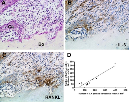
Figure 2.
Distribution of RANKL-positive and IL-6-positive cells at the interface of the tumor front and resorbing bone in human gingival SCC. A: Histological analysis of tumor-bone interface. Ca, cancer cells; Bo, bone. H&E staining. B: Distribution of IL-6-positive cells. C: Distribution of RANKL-positive cells. Cells stained brown in B and C represent positive cells for each antibody. Original magnification, ×200. D: Correlation between the number of RANKL-positive and IL-6-positive fibroblastic cells at the tumor-bone interface in 10 human gingival SCCs; a positive correlation was noted (r = 0.935926, Y = 0.8115X − 28.644, P < 0.0001).

