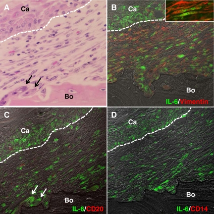
Figure 3.
Characterization of IL-6-positive cells at the interface of the tumor front and resorbing bone in human gingival SCC. A: Histology of the tumor-bone interface. H&E staining. B: Dual immunohistochemical analysis for IL-6 (green) and vimentin (red). Inset: High magnification image of the fibroblastic cells dual positive for IL-6 and vimentin. C: Dual staining for IL-6 (green) and CD20 (red). D: Dual staining for IL-6 (green) and CD14 (red). A and C are the same section; the section was stained with H&E (A) after pictures were taken for immunohistochemical analysis in (C). White dotted lines indicate interfaces of cancer nest and stroma. Arrows in A and C indicate osteoclasts. Ca, cancer cells, Bo, Bone. Original magnification: ×400 (A–D); ×1600 (inset in A).

