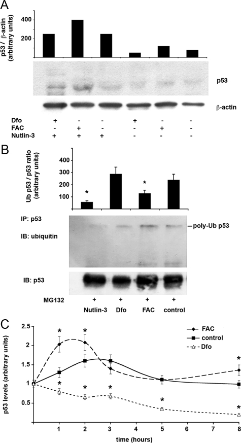
Figure 5.
Effect of iron status on p53 expression is mediated through MDM2. A: Effect of Nutlin-3 on iron mediated regulation of p53 levels. The effect of manipulation of iron status with FAC and Dfo on p53 is compared in cells exposed or not to Nutlin-3. Human monocytes isolated from a healthy subject with normal iron parameters and negative for HFE mutations were cultured for 24 hours after removal of lymphocytes in the presence or in the absence of 25 μmol/L Nutlin-3, 100 μmol/L Dfo, and 150 μmol/L FAC. β-actin is shown as a loading control. p53 levels normalized for β-actin, as detected by densitometry, are shown in the upper part of the figure. Results are representative of two independent experiments. B: Effect of iron status on p53 polyubiquitination during inhibition of proteasomal activity. Human monocytes isolated from a healthy subject with normal iron parameters and negative for HFE mutations were cultured for 24 hours in the presence or not of 25 μmol/L Nutlin-3, 100 μmol/L Dfo, and 150 μmol/L FAC in the presence of the proteasomal inhibitor MG132. Proteins were immunoprecipitated with p53 antibody and pull-downs blotted with antibodies against ubiquitin (upper panel) and p53 (lower panel). The histogram shows the average levels of ubiquitylated/total p53 as determined by densitometric analysis of two independent experiments. *P < 0.05. C: Iron status affects p53 degradation kinetics. Human monocytes isolated from a healthy subject with normal iron parameters and negative for HFE mutations were cultured for 24 hours after removal of lymphocytes in the presence or not of 120 μmol/L Dfo, and 150 μmol/L FAC for 24 hours and then treated with CHX 40 μg/ml for 1 to 8 hours. Western blot analysis and densitometric analysis were performed for p53 levels, which were normalized for β-actin. Thus, relative expression >1 compared with baseline means that protein degradation was slower, whereas relative expression <1 means that protein degradation was faster than that of β-actin. Results represent means and SDs of two independent experiments. *P < 0.05 compared with controls.

