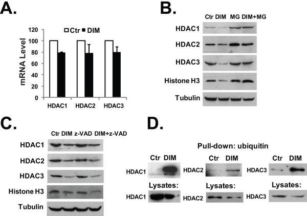
Fig. 2.
DIM induces proteasome-mediated degradation of the HDACs. (A) HT-29 cells were treated with 40 μM DIM for 24 hours, total RNA was isolated and real-time PCR analysis was done as described in Materials and Methods. (B) HT-29 cells were treated with 40 μM DIM, 10 μM of MG-132, or 40 μM DIM plus 10 μM of MG-132 for 24h. Western blotting was performed with the indicated antibodies. (C) HT-29 cells were treated with 40 μM DIM, 20 μM of z-VAD, or 40 μM DIM plus 20 μM of z-VAD for 24h. Western blotting was performed with the indicated antibodies. (D) HCT-116 cells were transfected with plasmids to express Flag-tagged HDAC proteins. 24h after transfection, cells were treated with 40 μM DIM for additional 24 hours. Cell lysates in RIPA buffer were collected and ubiquitin-modified proteins were isolated as described in Materials and Methods, followed by western blot analysis. The experiments have been repeated three times.

