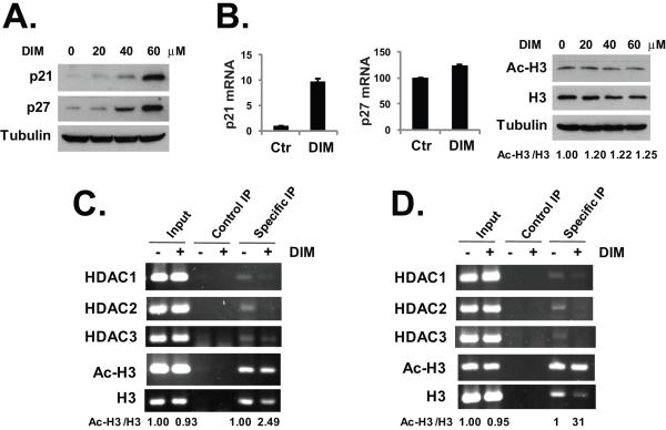
Fig. 3.
DIM activates p21 and p27 expression. (A) HT-29 cells were treated with various doses of DIM for 24 hours. Western blotting was performed with the indicated antibodies. (B) HT-29 cells were treated with 40 μM DIM for 24 hours, total RNA was isolated and real-time PCR analysis was done as described in Materials and Methods. For western blot analysis, HT-29 cells were treated with various doses of DIM for 24 hours. Western blotting was performed with an anti-acetylated H3 and anti-H3 antibodies and anti-tubulin antibody. (C) HT-29 cells were treated with 40 μM DIM for 24 hours. ChIP assay was performed as described in Materials and Methods, using primers specific for the p21 promoter and the indicated antibodies. (D) HT-29 cells were treated with 40 μM DIM for 24 hours. ChIP assay was performed as described in Materials and Methods, using primers specific for p27 promoter and the indicated antibodies. Relative protein levels and DNA band signals were quantified and shown under the gels. The experiments have been repeated three times.

