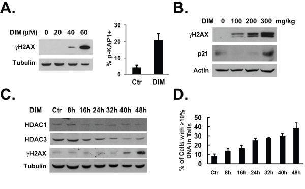
Fig. 5.
DIM induces DNA damage. (A) Left, HT-29 cells were treated with various doses of DIM for 24 hours. Western blotting was done using indicated antibodies. Right, HT-29 cells were cultured in chamber slides and treated with 40 μM of DIM for 48 hours. Immunofluoresence staining with anti-phospho-KAP1 antibody was done as described in Materials and Methods. The percentage of cells that are positive for KAP1 phosphorylation was determined after counting approximately 100 cells in each experiment. The average percentage of three independent experiments was shown. (B) Nude mice treatment was described in Fig. 1C. Tumor samples were analyzed by western blotting with indicated antibodies. (C) HT-29 cells were treated with 40 μM of DIM for various lengths of time as indicated. Western blotting was done using the indicated antibodies. (D) HT-29 cells were treated with 40 μM of DIM for various lengths of time as indicated. Comet assay was performed as described in Materials and Methods. The average results from two independent experiments were shown.

