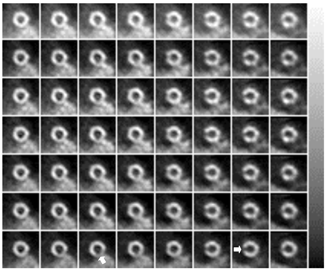
Figure 9.
Reconstructed short-axis Tl images for the anthropomorphic phantom experiment for the cases of (from top to bottom): no cross-talk compensation, TWC, SimMBC without x-ray model, SimMBC with x-ray model, SeqMBC without x-ray model, SeqMBC with x-ray model, and ‘no cross-talk’ image. The approximate positions of the lesions are indicated on the ‘no cross-talk’ images. All images were displayed using the same grayscale, shown at the right.

