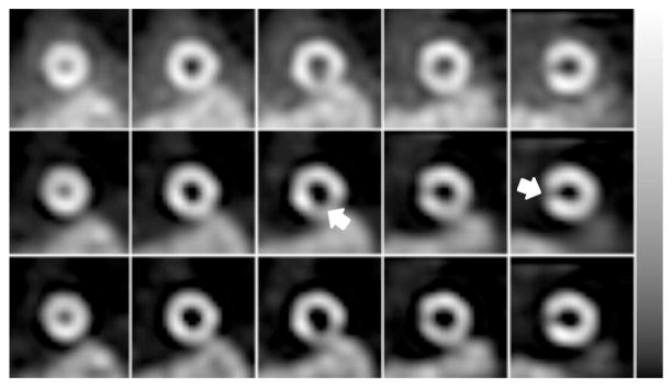
Figure 10.
Every other short-axis slice reconstructed for the Tl-201 phantom experiment using No Scatter Compensation (top row); Full RBSC (middle row); and Full/Intermittent RBSC, 2× (bottom row). The images of each row have been displayed using the grayscale shown at the right. The phantom had two cold lesions (arrows) in the myocardium—one in the basal septal wall, and another in the inferior mid-ventricular wall.

