Figure 3.
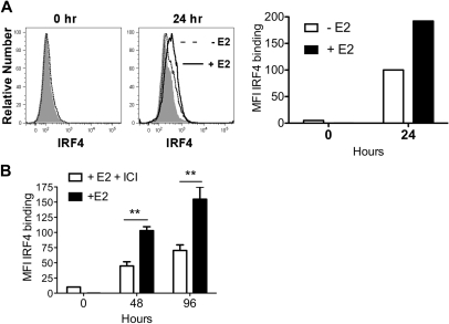
E2 exposure increases the amount of IRF4 protein in differentiating MPs 2-fold by 24 hours. MPs were stimulated with GM-CSF in hormone-deficient medium plus 1nM E2 or vehicle. (A) Shown is the binding of anti-IRF4 Ab to differentiating MPs in the absence (dotted line) or presence (solid line) of E2 at 24 hours. The shaded histogram indicates the binding of anti-IRF4 Ab preincubated with blocking peptide. The mean fluorescence intensity (MFI; with value for anti-IRF4 Ab bound to blocking peptide subtracted) of anti-IRF4 binding at 0 and 24 hours is plotted. Data are representative of 2 experiments. (B) MPs were stimulated with GM-CSF in the presence of E2 or E2 plus ICI 182,780 (100nM). The amount of intracellular IRF4 protein at 48 and 96 hours was assessed using flow cytometry. Shown are the MFI values (mean ± SD) of anti-IRF4 binding (with value for anti-IRF4 Ab bound to blocking peptide subtracted) of cells in triplicate cultures. **P < .01, n = 3.

