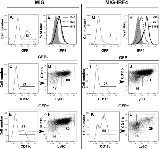
Figure 6.
Enforced IRF4 expression directs the development of the ERα/E2-dependent CD11bint Ly6C− DC subset in the absence of ERα signaling. Lin− ERα−/− BM cells were transduced with MiG-GFP (A-F) or MiG-IRF4-GFP (G-L). Transduced cells were incubated in GM-CSF for 5 days before assessment of DC subsets by flow cytometry. IRF4 expression was assessed in GFP− (dotted line) and GFP+ (solid line) fractions in (A-B) MiG-GFP– or (G-H) MiG-IRF4-GFP– transduced cells. The MFI of anti-IRF4 binding to GFP+ and GFP− cells is indicated in panels B and H. The shaded histogram indicates the binding of anti-IRF4 Ab precincubated with blocking peptide. In MiG-GFP–transduced cell cultures, CD11c+ DCs in the GFP− (C) and GFP+ (E) cell fractions are primarily CD11bhi Ly6C+ (panels D and F gated on CD11c+ cells shown in panels C and E). The percentage of cells with the bar or box gates is indicated. In MiG-IRF4-GFP–transduced cultures, CD11c+ DCs in the GFP− cell fraction (I) are also primarily CD11bhi Ly6C+ (J, gated on CD11c+ cells shown in panel I). In contrast, the percentage of CD11c+ DCs in the GFP+ fraction (K) is significantly increased, and the majority of DCs is CD11bint Ly6C− (L, gated on CD11c+ cells shown in panel K). Data are representative of 4 independent experiments.

