



Abstract
We draw on the theory of allostasis to develop an integrative model of the current stress process that highlights the brain as a dynamically adapting interface between the changing environment and the biological self. We review evidence that the core emotional regions of the brain constitute the primary mediator of the well-established association between stress and health, as well as the neural focus of “wear and tear” due to ongoing adaptation. This mediation, in turn, allows us to model the interplay over time between context, current stressor exposure, internal regulation of bodily processes, and health outcomes. We illustrate how this approach facilitates the integration of current findings in human neuroscience and genetics with key constructs from stress models from the social and life sciences, with implications for future research and the design of interventions targeting individuals at risk.
Keywords: stress, brain, allostasis, allostatic load, genes
Introduction
“There is growing evidence that multilevel analyses spanning neural and social perspectives can foster comprehensive accounts of cognition, emotion, behavior, and health. This is in part because the social environment shapes neural structures and processes and vice versa” (Cacioppo, Ernst et al., 2000, p. 152).
This review and synthesis covers broad themes and key findings across numerous areas of stress research to develop a new framework for converging biomedical and psychosocial models of the stress process. This effort expands on theory that identifies the brain as the central mediator of ongoing systemwide physiological adjustment to environmental challenge (McEwen, 2004, 2007; Schulkin, 2003; Sterling & Eyer, 1988; Sterling, 2004). This process has been termed allostasis (Sterling & Eyer, 1988), a major revision (McEwen 2004) or replacement (Sterling, 2004) of the classical theory of homeostasis. Exploiting the central role of the brain in allostasis, we integrate new research in human neuroscience and genetics with key elements of stress models from the social and life sciences to develop a model of the interplay over time between current stressor exposure (in its physical, social, and cultural context), the internal regulation of bodily processes, and health outcomes. Integration of social and life science research perspectives simultaneously constrains models of the stress process at all levels of analysis and prompts their expansion to include new methods, mechanisms, and objectives, with implications for the development of a research agenda and the design of interventions targeting individuals at risk.
In the expanding research utilizing allostatis as a working model of the relationship between environmental challenge and physiological outcomes, the focus has been on the effect of prior stressors (e.g., McEwen & Seeman, 2003; Singer, Ryff, & Seeman, 2004). We expand on these earlier models to include response to a current stressor, which we term allostatic accommodation. Modeling the ongoing stress process requires a much higher level of specificity about the underlying mechanisms of allostasis. To this end, we argue that the core emotional regions of the brain1 serve as the primary mediator of physiological and behavioral response to a current stressor and that other physiological and behavioral change in the face of adversity is secondary to this central mediation. It follows that the core emotional regions of the brain will be the first and primary sites to show evidence of long-term “wear and tear” (McEwen & Seeman, 1999; McEwen & Stellar, 1993) as a part of the cost of physiological accommodation to environmental demand. Modeling the ongoing stress process also demands a higher level of specificity about the stressors themselves and their relationship to proximal and distal risks and resources in the environment. Integration of allostasis with recent findings in neuroscience and dynamic systems theories regarding the context of stress gives us a new theoretical platform for incorporating biology into the social psychological study of stress.
The result of this effort is a model delineating the individual’s response to a current stressor. This model reveals systematic sources of variation in the stress response itself that render it mandatory to explore the full set of associations between personal stress history, the stressor in context, brain, and body to best understand health outcomes. Within this allostatic model, an organism’s ability to “achieve stability through change” (p. 171, McEwen, 1998) represents ongoing development. Establishing ongoing development within the stress response itself focuses a new lens on the etiology of acquired vulnerability and resistance to stress across the lifespan. This prompts specific predictions about the impact of accumulating life stress within an array of research domains, with suggestions for future research.
This effort is prompted by the observation that the social and biological approaches to stress research had, over many years, diverged into largely parallel structures with independent academic research traditions, methodologies, and literature (Cacioppo, Berntson, Sheridan, & McClintock, 2000; Magnusson, 1995; Mason, 1971). These research traditions are now beginning to converge again, with notable interfaces arising in a number of disciplines, including human development (e.g., Cicchetti & Rogosch, 2001; Granger, Hood, Dreschel, Sergeant, & Likos, 2001; Gunnar & Vasquez, 2001; Susman, Schmeelk, Ponirakis, & Gariepy, 2001) and gerontology (e.g. Karlamangala, Singer, McEwen, Rowe & Seeman, 2002; Seeman, Crimmins, Huang, Singer, Bucur, Gruenwald et al., 2004), and emerging in new disciplines such as biosocial and public health epidemiology (Geronimus, Hicken, Keene, & Bound, 2006; Hubbs-Tate, Nation, Krebs, & Bellinger, 2006; Krieger, 2001) and social neuroscience (e.g., Caccioppo et al, 2007). In addition, new neuroimaging techniques are driving a renaissance in brain-body medicine (e.g., Lane et al, 2009; Lane & Wager, 2009). This, together with research involving clinical outcomes of stress-related disorders (mechanisms, treatment, epidemiology), has also served as an important interface between the fields of biomedical and psychosocial stress research (e.g., Brown, 2002; Caspi, et al., 2002; Cohen, Kessler, & Gordon, 1995; Gallo & Matthews, 2003; Glaser, Kiecolt-Glaser, Malarkey, & Sheridan, 1998; Moffit, Caspi, & Rutter, 2006; Rutter, 1994). Notably, the convergence of the social and biological research traditions has lacked a common model for specifying the stress process. The focus of the present paper is the development of such a model.
The model we propose here is potentially of use to stress researchers in the life sciences who are faced with the need to broaden their research agendas to include the impact of social/environmental influences on outcomes in a range of fields, including neuroscience, genetics, neuroendocrinology, medicine, and immunology. For example, identification of the core emotional systems of the brain as the neural foci of response and adaptation to stressor exposure is important because these will be the first and primary sites to show evidence of long-term “wear and tear” (McEwen & Seeman, 1999; McEwen & Stellar, 1993) as a part of the cost of physiological accommodation to environmental demand, including social context. Thus, this model raises questions regarding long-term stress-related plasticity and damage in the healthy human brain, with implications for present and future work in social neuroscience that we will discuss later. We also note the existence of an inverted U-shaped relationship between stressor exposure and adaptation, such that small-to-moderate amounts of stressor exposure (often termed stimulation or challenge) lead to increased health and improved physiological and mental function, and high levels of stressor exposure are related to negative health outcomes (see McEwen, 2002, for a review). For example, profound early deprivation of environmental stimulation is known to be detrimental to brain development, cognitive function, and socioemotional adaptation (Ames, 1997; Rutter, 1998) and progressive increases in environmental stimulation (up to a point) are associated with increases in neurocognitive function (Farah et al., in press) and other health outcomes (McEwen, 2002). While we will discuss this allostatic model primarily in terms of the “high-stress” side of this inverted U-shaped curve (where the majority of research on negative health outcomes has been concentrated), we will also use this model to articulate the difference between a helpful challenge and a harmful stressor, as well as who may be most stress-vulnerable (or resilient), when, and why. These are relatively new questions for social neuroscientists but they will resonate with stress researchers in the social sciences, for whom these are long-standing concerns. In addition, a common theoretical platform would be of use to social scientists who are faced with a rapidly advancing array of discoveries in the life sciences and who need to assess the potential usefulness of these advances in furthering psychosocial research agendas. While the rich literature on human stress and coping in the social sciences can serve to direct biological and medical research onto new and exciting ground, biological stress research (and human neuroscience in particular) can serve as an empirical testing ground for psychosocial theory and a means to refine the scope and methods of intervention research.
This paper is organized as follows: In the first section, we take the opportunity to understand the historical points of divergence and current points of intersection between stress research in the social and life sciences. Then we discuss the fundamentals of allostasis and how it functions in the integration of these research tranditions. This review will allow readers with diverse background to have a common understanding of the foundations of our model. In the second section, we briefly review the relevant brain anatomy and build our model of the allostatic response to a current stressor in context, with the brain as the master controller of this physiological adjustment and the primary regulator of allostasis. This is the point of transition in this work from a review of prior work to the development of our proposed, integrated model. We then model the current stress process from the perception of proximal and distal stressors in context, through central and peripheral accommodation as modified by prior allostatic load, with mental and physical health as primary outcomes. We conclude our model-building by bringing time and genetics in to the model. Throughout this section, we review the current evidence that stress-related neural changes in the healthy human brain play a key role in the well-established association between stress exposure and long-term decreases in well-being and health and suggest points for future research. In the final section, we summarize these points for future research and draw from our new conceptual framework to make predictions based on this model. Finally, we discuss implications of the model for new research and interventions targeted at individuals at risk.
Common Origins: Psychosocial and Biological Models of the Stress Process
Walter Cannon and homeostasis
Stress research in the social and life sciences shares a common foundation in the work of Walter Cannon (Cannon, 1932) and Hans Selye (Selye, 1956). At the beginning of the 20th century, Harvard physiologist Walter Cannon expanded upon Claude Bernard’s views of a flexibly stable milieu interieur (Bernard, 1878: also see Goldstein, 1995a) in his investigation of the response of the sympathetic-adrenal medullary system to emergency situations. This system swiftly mobilizes the body’s energy resources by increasing epinephrine (adrenaline), which in turn increases blood pressure, heart rate, and blood sugar, as well as hastening blood coagulation, clearing fatigue products from muscles, and decreasing digestion (Cannon, 1920). In Cannon’s view, these processes occurred locally in the body, independent of central nervous system (CNS) control. He used the term homeostasis to refer to the way this array of independent physiological systems works together to re-establish initial conditions when the system is perturbed (Cannon, 1932).
Selye, the GAS, and the “unknown mediator”
Hans Selye demonstrated that an organism has an adaptive response to adversity that includes both Cannon’s sympathetic (adrenaline-driven) responses plus the actions of hormones from the pituitary gland, which globally affect the major organs of the body in indirect but important ways (e.g., Selye, 1956). Selye proposed the existence of a generalized physiological syndrome that occurs in response to a great diversity of threats to the integrity of the organism (e.g., Selye, 1956). The syndrome, which he called the General Adaptation Syndrome or GAS (Selye, 1956), included three stages of sequelae to stressor exposure. The alarm stage is best characterized by dramatically increased activity of the hypothalamic-pituitary-adrenal (HPA) axis. The HPA axis starts in the brain, where an area of the hypothalamus (the paraventricular nucleus) is activated by perceived adversity and begins to produce corticotropin releasing hormone (CRH, also called corticotropin releasing factor, CRF). These, in turn, activate the anterior pituitary to produce adrenocorticotropin releasing hormone (ACTH), which causes the adrenal cortex to produce corticosteroids (cortisol, in humans). Corticosteroids, in turn, act broadly and relatively slowly to mobilize the body’s energy resources. In the resistance stage of the GAS, the organism’s biological responses to the stressor achieve a steady state and overt symptoms of stress often are reduced or disappear. Eventually, however, the organism enters the third stage of the GAS, which Selye called exhaustion. If a stressor continues and physiological defenses are depleted, then the organism’s symptoms reappear during the exhaustion stage. If there is no relief, death follows.
Selye adopted the term “stress” to describe this overall phenomenon and re-defined it to mean “that which stimulates the GAS response” (Selye, 1956, p. 54). He tested the universality of all three stages of the GAS across a very broad range of severe stressors (e.g., physical trauma, burns, heat, cold, electroshock, infection) and suggested these stressors shared the common trait of being noxious (Selye, 1950). His writing included discussion of a hypothetical neural or endocrine “common mediator” that lies between application of the noxious stressor and the GAS response (Selye, 1950, p. 105). This unknown “common mediator” would serve to receive and integrate local inputs and then transmit generalized messages to all parts of the body to activate the GAS response, including facilitating changes in regulatory (i.e., homeostatic) set-points during the stage of resistance (e.g., Selye, 1950, 1956).
Although Selye’s research drew into question the stability of the internal milieu (Bernard, 1878; Cannon, 1932), his work retained many of the omissions inherent in the original conceptualization of homeostasis, including inattention to the significance of psychological state, information processing, life history, and environmental context beyond the specific stressor (Mason, 1975b; Burchfield, Woods, & Elich, 1980; Toates, 1995). Importantly, he also never identified the “common mediator” that facilitated changes in homeostatic set-points in response to the environment. This removed consideration of many of the central questions of the social sciences from biological models of the stress process and foreshadowed the divergence of stress research in the social and life sciences over subsequent decades.
Diverging from Cannon and Selye
Selye’s claims for the scope of application of the GAS were sweeping (e.g., Selye, 1956) and his work was highly provocative. It prompted thousands of articles across the fields of medicine, biology, psychology, and sociology (Goldstein, 1995b; Mason, 1975b). Initially, and with Selye’s encouragement (e.g., Selye, 1956), early stress researchers across many domains invoked Selye’s concept of the nonspecificity of stressors to justify application of the GAS to the entire range of systemic (e.g., heat, cold, infection, hemorrhage) and psychosocial stressors (both positive and negative, e.g., the Schedule of Recent Experiences; Hawkins, Davies, & Holmes, 1957). The psychosocial stress literature of this era demonstrated a reliable association between life events and both psychological distress and medical disorder (Turner & Wheaton, 1995). This research, in turn, engendered an increasing amount of critical commentary pointing out that the size of these associations was persistently small (e.g., Thoits, 1983). It was noted that more explicit measurement of the chronicity, as well as the level of change or threat posed by a given stressor often improved the amount of variance explained in the outcomes, (e.g., Brown & Harris, 1978; Dohrenwend, Ashkenasy, Krasnoff, & Dohrenwend, 1978; Holmes & Rahe, 1967; Pearlin & Schooler, 1978; Wheaton, 1999b) and it became clear that changes in environmental context over time made significant independent contributions to the outcomes of stressor exposure (e.g., Baltes & Baltes, 1990; Furstenberg, Brooks-Gunn, & Morgan, 1987; Hobfoll, Johnson, Ennis, & Jackson, 2003), Holahan, Moos, Holahan, & Cronkite, 1999; Lazarus, 1993a; 1999; Masten et al., 1988; Rutter, 1979; Sameroff, Seifer, Barocas, Zax, & Greenspan, 1987). Importantly, individual differences in cognitive and emotional responses to both stressor and context were found to be key factors in determining outcomes (e.g., anticipation, appraisal, coping, learning, and other types of information processing: Foa & Kozak, 1986; Lazarus, 1991; Hobfoll, 1989; Holahan, Moos, Holahan, & Cronkite, 2000; Ironson et al., 2000; Siegal & Allan, 1998; Sterling & Eyer, 1988; Stone, 1995; Toates, 1995; Tolin & Foa, 2002; Wheaton, 1985). Thus, within the social sciences, the historical process of improving the power of psychosocial factors in predicting health outcomes has been one of more carefully specifying the accumulating negative impact of different stressors within a changing environmental context, combined with acknowledgment of individual differences in response and adaptation over the lifecourse (e.g., Baum, Garofalo, & Yali, 1999; House et al., 1992; Singer & Ryff, 1999). Taken together, this body of work has stretched the psychosocial requirements for models of the stress process well beyond the classical homeostatic perspective or the GAS.
Parallel streams of discovery
Within biomedical research, observations of the influence of the brain and the environment on the stress response process were also prompting reconsideration of classical homeostasis (for a review, see Schulkin, 2003) and Selye’s GAS (Mason, 1971, 1975b; Pacak & Palkovits, 2001). Mason (1971, 1975a) documented substantial variation in elements of the GAS response as a function of the situation, the individual, and the individual’s history, thus raising questions about the specificity of the “nonspecific” GAS response (also see Pacak & Palkovits, 2001). He also discovered that whenever it was possible to substantially reduce or remove the noxious “psychological concomitants” of the stressor, then there was no GAS response (Mason, 1971, 1975a, p. 326); “The knowledge that the psyche is superimposed upon the humoral machinery for endocrine regulation drastically complicates our whole view …” (Mason, 1975a, p. 177). Thus, the biomedical stream of stress research also found itself in need of a model that included appraisal, cognition, and emotional state as primary elements of the stress process (Frankenhaeuer, 1979; Moore-Ede, 1986; Weinberg & Levine, 1979).
This work set the stage for Sterling and Eyer’s (1988) presentation of allostasis, which proposed that the central nervous system (CNS) exerts control over all physiological regulatory responses to environmental demand in the alert, intact organism. They argued that control of the stress response by the CNS allows the regulatory set-points of the organism to vary in response to environmental demand (Sterling & Eyer, 1988), thus placing the brain squarely in the role of Selye’s “common mediator” between the environment and physiological response (Selye, 1950). This work went largely unnoticed at the time of its publication. Three years later, Levine and Ursin (1991) proposed that the sensory input from all types of stressors is gated through the brain before affecting any other physiological regulatory response, and that this input is modified by expectation and evaluation. They argued that this is true even of stressors that do not appear overtly “psychological,” such as cold exposure and tissue damage, because novelty, expectation, and efforts to avoid noxious stimuli will all activate brain responses (Levine & Ursin, 1991). Soon after, Chrousos and Gold (1992) and Goldstein (1995a, b) independently proposed a model in which homeostasis “resets” itself in response to stress exposure and Goldstein (1995b, p. 41) argued that “distress invariably resets homeostasis.” Thus, by the end of the 20th century, it was clear that homeostasis required revision to account for the influence of environment and/or CNS influence on physiological regulation.
None of these modifications of homeostatic theory addressed the accumulating cost of dynamic, ongoing physiological accommodation to environmental challenge over the lifespan. This prompted McEwen and Stellar to introduce the concept of allostatic load (McEwen & Stellar, 1993), the physiological cost of making long-term adaptive shifts across a broad range of systems in order to match internal functioning to environmental demand. With the inclusion of allostatic load as a key element of the theory, allostasis became the most comprehensive account of regulatory accommodation to environmental demands and accumulated physiological cost over time. The adaptation and dissemination of allostatic theory by McEwen and an expanding cohort of colleagues (e.g., McEwen, 1998; McEwen, 2000a; McEwen, 2003a, 2003b, 2003c, McEwen & Seeman, 1999; McEwen & Seeman, 2003; McEwen & Stellar, 1993; Schulkin, Gold, & McEwen, 1998; Schulkin, McEwen, & Gold, 1994; Schulkin, 2003, 2004; Singer, Ryff, & Seeman, 2004) is lending allostasis the status of “a new conceptual framework” for the study of stress (Schulkin et al., 1998, p. 220), with advantages for research in both the social and life sciences.
Allostasis
Advantages for modeling the stress process
Because homeostatic responses operate locally, they can be (and typically have been) studied in organs and tissues that have been isolated from the body. Thus, for much of the last century, the biological response of the body to stressors has been viewed as largely independent of the brain. When Sterling and Eyer (1988; see also Sterling & Eyer, 1981; Sterling, 2004) pointed out that homeostatic models do not provide adequate explanation for the internal regulation of an unanesthetized animal that is experiencing a variety of behavioral states in response to an ever-changing environment, they argued that essentially all physiological parameters2 covary with behavioral state and that behavioral state is identified with CNS activity (or “states of the brain”, Schulkin, 2003, p. 12). Thus, the brain has overriding regulatory power that supercedes the local effects of homeostasis in the conscious organism. Sterling and Eyer (1988) note that this is a more complex system of regulation than classical homeostasis but it has several advantages for stress researchers in both the social and life sciences:
Allostasis allows ongoing evaluation of the match between internal resources and external demands.
Allostasis permits the organism to produce physiological adjustments in advance of need through anticipatory arousal.
Allostasis allows the organism to adapt to circumstances over time.
Allostasis predicts the responses of an alert, intact organism existing in a changing ecological framework.
This is illustrated in the etiology of chronic high blood pressure. Cannon (1920) and Selye (1956) both developed their theories by studying animals that were subjected to varying levels of environmental stressor exposure, resulting in acute or semichronic arousal. In these situations, blood pressure rises during arousal and falls afterwards. Sterling and Eyer (1988) pointed out that neither the classical view of homeostasis nor the general adaptation syndrome (GAS) provides a mechanism by which blood pressure fluctuates markedly throughout the day, nor a mechanism to maintain elevated blood pressure once a stressor is removed (Sterling, 2004). Consistent with an allostatic model, an animal’s blood pressure typically does not fall when faced with arousal over a sustained period (Forsyth, 1969). In short, the system is responding to “anticipated demand” (Sterling, 2004, p. 24), as the aroused brain activates the sympathetic and hormonal systems. These increase cardiac output, blood volume, sodium appetite (and thus sodium intake), and vascular constriction to temporarily raise blood pressure. When arousal becomes chronic, the body adapts by thickening arteriolar smooth muscle and increasing the vascular wall-to-lumen ratio so that they are more effective in maintaining a higher blood pressure (Boulos & Rosenwasser, 2004; Sterling, 2004). For such individuals, even under conditions of maximum relaxation, blood pressure does not decrease to the previous low point. This is because blood pressure is maintained by vascular resistance such that a higher blood pressure is needed to maintain the same blood flow (Boulos & Rosenwasser, 2004; Folkow & Neil, 1971; Lund-Johanson, 1984). The vascular system, under the direction of the aroused brain, has adjusted its parameters to meet the demands of the chronically stressful environment. This is an adaptation that is rarely reversed, even when the chronic stressor is removed (probably because of hormones that feed back to the brain to maintain this efficiently-responding system: Sterling, 2004).
Thus, when a physiological system is activated by a current stressor, it accommodates by adjusting its parameters within a range of functioning (allostatic accommodation). “Different circumstances demand different homeostatic set points” (Sapolsky, 1998, p. 7). These changes will have cascading effects in other systems, producing organism-wide accommodation to the environment. Thus, in response to a stressor, the organism is not struggling to get its homeostatic systems back into their initial balance. Instead, it is making wide-ranging physiological changes in order to find a new homeostasis that better fits the circumstances (McEwen, 1998). By identifying the brain as the central mediator of allostatic accommodation to a stressor, Sterling and Eyer’s (1988) theory of allostasis accounts for the effects of anticipatory arousal, appraisal, coping, learning, and memory on physiological regulation under adversity and subsequent effects on health.
Notably, under conditions of repeated or chronic stress, this system-wide accommodation can combine with the physiological tendency for the body to create “fixed automatisms” (Sterling & Eyer, 1988, p. 641) and cause the lasting cardiovascular alterations associated with high blood pressure, as well as a range of other possible changes, such as alterations in the concentration and receptor densities of key hormones (e.g., Blanchard, McKittrick, & Blanchard, 2001; Yehuda, 1997) or irreversible activation and/or downregulation of the transcription of specific genes (e.g., Levine, 2001; Valentino & Van Bockstaele, 2004). Implicit in this analysis is the notion that allostatic accommodation over time also results in“wear and tear” in the very systems that are experiencing adaptation to meet the environmental challenge (McEwen, 1998).
Allostatic load: The impact of prior stressors
Rutter has referred to the effects of prior stressors as the “long-term carry-forward of the sequelae of stress and adversity” (Rutter, 1994, p. 373) and argued that they must be accounted for in any formulation of the stress process. In allostasis, the new balance of system parameters that follows stressor exposure comes at a physiological cost that McEwen and Stellar (1993) have called allostatic load. This cost can occur through the adoption of Sterling and Eyer’s “fixed automatisms” (1988. p. 641), i.e., those small and large physiological changes that do not revert to the way they were when the challenge has passed. Such changes may be helpful in the short term (as with the increases in blood pressure discussed above) but may have negative long-term consequences for the organism (e.g., increased wear-and-tear on the heart). This cost can also come through damage due to overproduction of the neurochemicals involved in the stress response, some of which are toxic. For example, persistent high concentrations of cortisol can cause damage to regions of the hippocampus (e.g., Bremner et al., 1995; Gurvits et al., 1996; Sapolsky, 1984; Sapolsky et al., 1986; Uno, Tarara, Else, Suleman, & Sapolsky, 1989) and inhibit neurogenesis in this region (Gould, McEwen, Tanapat, Galea, & Fuchs, 1997), both of which potentially interfere with cognition and future adaptation to stressors. Allostatic load can also occur through exhaustion of stress response systems, as can occur in the immune system. This can result in compromised immunocompetence, which is related to higher levels of infection and vulnerability to cancer (Cohen, Tyrrell, & Smith, 1991; Sapolsky, 1998; Sapolsky & Donnelly, 1985). Increased load can also come through the inability to activate a particular stress response system, in which case other stress responses over-respond (McEwen, 1998).
Allostatic load can be negligible, resulting in a symptom-free organism that is well adapted to living with a particular stressor. This is analogous to remaining within the “elastic limit” of the human material in the “engineering” model of stress described by Cannon (1935). Human resilience under duress (e.g., Freitas & Downey, 1998; Garmezy, 1996; Glantz & Sloboda, 1999; Kaplan, 1999; Rutter, 1990) may be seen as resistance to the acquisition of allostatic load, which may help concretize this important but elusive concept (Glantz & Sloboda, 1999; Tarter & Vanyukov, 1999). Load can accumulate from daily low levels of stress in the environment as well as from discrete life events, as observed in the effects of social hierarchies on physical health in nonhuman primates (e.g., Kaplan, Adams, Anthony, Morgan, Manuck & Clarkson, 1995; Kaplan, Manuck, Clarkson, Lusso, & Taub, 1982; Kaplan, Manuck, Clarkson, Lusso, Taub, & Miller, 1983; Manuck, Marsland, Kaplan, & Williams, 1995) and humans (e.g., the Whitehall studies: Marmot, Shipley & Rose, 1984; Marmot, Kogevinas & Elston, 1987; Marmot, 1994). On the other end of the stressor spectrum, allostatic load can be massive enough to produce symptoms that are severe, or even fatal, as in Selye’s “exhaustion” phase (Selye, 1956). The concepts of allostasis and allostatic load allow for dynamic change in the adaptive capacity of the organism and they highlight individual differences in ability to withstand stressors (which can be related to Antonovsky’s [1974] early concept of “homeostatic flexibility”). By defining this accumulating cost of physiological adaptation and outlining some of the biomarkers by which it might be identified, McEwen and Stellar (1993) provided a conceptual framework to explain the accumulated effects of prior experience on physical and mental health (e.g., Boulos & Rosenwasser, 2004; Heim et al, 2000; Levine, 2001; Mason, 1971, 1975a; Resnick, Yehuda, Pitman, & Foy, 1995).
Implications for an integrative model
In sum, history suggests that the coherent integration of social and biological approaches to stress research has been hampered by the traditional biological representations of the stress process. In particular, many of the fundamental findings of psychosocial stress research have been incongruent with the classical biological paradigm of homeostasis, which has been the dominant model of physiological regulation for the past 100 years (Schulkin, 2003). The classical view of homeostasis maintains that the physiological parameters of the body have an ideal set-point under the control of local biological mechanisms (Cannon, 1932), i.e., these parameters can and will return to equilibrium after perturbation without the need for a central mediator. Classical homeostasis does not easily reconcile with the observation that most of these physiological set points vary as a function of environmental context and psychological state (Sterling & Eyer, 1981, 1988, Sterling, 2004), resulting, for example, in the social stratification of incidence of chronic disease (House et al., 1992). Nor does homeostasis explain why increased social support ameliorates this risk (Seeman, Singer, Ryff, Love, & Levy-Storms, 2002; Singer & Ryff, 1999). Homeostasis alone does not account for the mitigating and exacerbating effects of emotional regulation, anticipation, appraisal, learning, and other types of information processing on the biological response to threat (Siegal & Allan, 1998; Sterling & Eyer, 1988: see also Lazarus, 1991; Hobfoll, 1989; Holahan, Moos, Holahan, & Cronkite, 2000; Ironson et al., 2000; Toates, 1995; Tolin & Foa, 2002; Schulkin, 2003). In addition, the classical view of homeostasis does not square with empirical evidence indicating that the stress response itself is not static: rather, it changes over time as a function of the life history of the individual (Levine, 2001; Liu et al., 1997; Mason, 1975a; McEwen & Stellar, 1993).
Although there has been recognition that the classical homeostatic perspective presents problems in a number of scientific domains (for reviews, see Schulkin, 2003; 2004), these issues have been especially pressing for social scientists. Social scientists typically study organisms that are intact, alert, and functioning in a changing ecological framework (as opposed to being observed dismantled and in vitro). Under these circumstances, the brain itself exerts executive control over local regulatory systems (Sterling & Eyer, 1988; Sterling, 2004), as we will discuss in detail below. With the central nervous system in charge, physiological system parameters do not necessarily return to their pre-perturbation state; instead, they co-vary with behavioral state to re-establishing new set-points that are better suited to challenges presented by the environment (McEwen, 1998; 2000a; 2003a and c, 2004; Sapolsky, 1998; Schulkin, McEwen, & Gold, 1994; Sterling & Eyer, 1988). This CNS-driven, continuous adjustment to ongoing environmental challenge is the keystone of our approach to modeling the current stress process.
Allostasis and the Brain
Overview
It is at this point that we transition from a review to a new synthesis of the emerging literature on the human neuroscience of the stress response and psychosocial perspectives on the context of stressors. To do so, we will review the relevant neural systems and present an integrative model of the stress process that incorporates current research in human neuroscience with key elements of stress models from the social and life sciences (see Cohen et al., 1995, for a review of the distinctions between the two). The concepts of allostasis and allostatic load allow this integration. The model itself constitutes the framework of our proposed bridge between the traditions of stress research in the social and life sciences. As discussed, the keystone of this model is identification of the brain as the primary mediator of allostasis, i.e., the primary point of interface between a given stressor in its physical and social context, and an individual’s physiological and behavioral accommodation to that stressor. We will emphasize here that the core emotional regions of the brain constitute the primary regulators of allostatic accommodation, to which all other physiological and behavioral accommodation is secondary. This follows from the recognition of these neural systems as the main organizing factor in the translation of environmental stimulus into behavioral and/or physiological response (e.g., LeDoux, 1996; Ohman & Mineka, 2001; Rosen & Schulkin, 1998). Identification of the neural foci of allostatic regulation is important because these will be the first and primary sites of physiological adaptation in the face of adversity, as well as the areas mostly likely to show evidence of allostatic load. Accrual of allostatic load in the brain will have consequences for psychosocial adaptation and health; identification of the specific circuitry that is most vulnerable to the acquisition of load will allow better understanding of the physiological and behavioral consequences of environmental adversity in humans, which will, in turn, allow more precision in intervention strategies to aid populations at risk. Thus, identification of the neural processes underlying the central regulation of allostasis (and the central acquisition of load) is our first step in bridging stress models in the social and life sciences.
Emotions and the brain
Emotions are central motivational states of the brain that underlie human behavioral adaptation to the environment (LeDoux, 1996; Ohman & Mineka, 2001; Panksepp & Miller, 1996; Rosen & Schulkin, 1998; Schulkin, 2003). The weight of current experimental evidence across multiple species suggests that control of emotional processing lies in the central nervous system. Further, this control is primarily localized within a limited number of brain regions that function together as a neural circuit or set of interconnected circuits (see below; e.g., Davidson et al., 2000; Gregg & Siegel, 2001; Ishai. Pessoa, Bilkle, & Ungerleider, 2004; LeDoux, 1995; LeDoux, 1996; Ohman & Mineka, 2001, Panksepp, 1998; Sanchez et al., 2001). The processing of emotional stimuli is regulated by neurotransmitters and neurohormones within this system, which in turn underlies emotion-related behavioral states and physiological response in systems external to the CNS (for reviews, see Gunnar & Quevedo, 2007; Schulkin, 2003; Salamone & Correa, 2002). These core emotional regions of the brain are highly interconnected with other neural systems that underlie perception, cognition, bodily representation, homeostasis, and behavior (see Pessoa, 2008, for a review); they serve as hubs in the processing of stimuli that have threat or reward value, which we will call emotional stimuli.
Two key areas in the neural systems associated with the processing of emotional stimuli are the amygdala and basal ganglia, both of which are highly evolved (Barton & Aggleton, 2000; Gerfen, 1992) structures or sets of structures near the base of the brain, below the neocortex. Meta-analyses indicate that these structures play a consistent (Murphy, Nimmo-Smith, & Lawrence, 2003; Phan, Wager, Taylor, & Lieberzon, 2002) and central (Wager, Phan, Lieberzon, & Taylor, 2003) role in the functional neuroanatomy of emotion in humans. This is supported by research with animal and humans, which also identifies the involvement of the extended amygdala3, hypothalamus, and a small number of medullary and brainstem nuclei as central to the processing of emotional stimuli (Armony & LeDoux, 1997; Koob, 2003; LeDoux, 1996; Panksepp, 1998).
The coordinated activity in these brain regions is seen in the interplay of the basic “approach” and “avoidance” mechanisms of the brain. In general, the basal ganglia (in particular, the nucleus accumbens) are associated with approach behavior, motivation, and positive incentive (e.g., Galvan et al., 2006; Haber, Kim, Mailly, & Calzavara, 2006), as well as playing important roles in motor and cognitive control (Casey, Tottenham, & Fossella, 2002). In contrast, the amygdala and extended amygdala are generally associated with fearful, anxious, and avoidance-related behavior (e.g., Davis, Walker, & Lee, 1997; LeDoux, 1996; Phelps, 2004, 2006). Yet these regions are highly interrelated. For example, the current reward value of a stimulus is encoded in the amygdala and orbitofrontal cortex (Gottfried, O’Doherty, & Dolan, 2003); conversely, the amygdala modulates the incentive values of rewards by controlling the amount of dopamine available to key regions in the basal ganglia (Phillips, Ahn, & Howland, 2003).
These regions are also highly interconnected with nearby brain areas that provide more complex representations of emotional stimuli and regulation and evaluation of emotional response (Bremner & Vermetten, 2001; Kaufman & Charney, 2001; Sanchez et al., 2001; also see supporting human neurimaging data, e.g., Dolcos, LeBar, & Cabeza, 2004; Kim et al., 2004; Wager et al., 2003; Wickers et al., 2003; Urry et al., 2006). The medial and ventromedial prefrontal cortices, located along the central and lower midline of the prefrontal cortex, are implicated in the representation and evaluation of the potential personal impact of a given stimulus. For example, functional imaging studies have found ventromedial (Phan et al., 2004) and medial (Lane, Fink, Chau, & Dolan, 1997; Phan et al., 2004) prefrontal cortex activation in response to tasks involving appraisal of the self-relatedness of emotional images; patients with damage to the ventromedial prefrontal cortex are observed to have difficulty generating an emotional response when recalling emotion-laden past experiences (e.g., death of a loved one: Bechara, Damasio, & Damasio, 2003). Interior to the medial and ventromedial prefrontal cortex is the affective subdivision of the anterior cingulate cortex, which is implicated in the regulation of emotional response (for a meta-analysis, see Bush, Liu, & Posner, 2000). This is closely connected with the ventromedial prefrontal cortex, insula, and hippocampus, as well as with the amygdala and the basal ganglia (Bush et al., 2000). The hippocampus, located adjacent to the amygdala, provides both representation of the context of an emotional stimulus (Phillips & LeDoux, 1992) and explicit knowledge of the pairing between a negative stimulus and an unconditioned one (Bechara et al., 1995), and serves a key role in the regulation of the HPA-axis (Sapolsky et al., 2000). Together, these brain regions form an interconnected neural circuit or set of circuits (often referred to as the limbic system, but see LeDoux, 1996, pp. 98–102) that serves to represent, regulate, and process emotional stimuli, with a widening array of neocortical areas being recruited with increasing task demand and complexity. The high level of structural and functional connectivity between these regions of the brain and the rest of the cortex allows these core emotional areas to function as hubs for recruiting and integrating a wide range of the brain’s computational resources in the processing of emotional stimuli (Pessoa, 2008; Stephan et al., 2000). Such stimuli effectively engage attention and are prioritized in the competition for processing resources in the brain (e.g., Anderson, 2005; Ohman, Flykt, & Esteves, 2001; Vuilleumier, Armony, Driver, & Dolan, 2001). This provides emotional stimuli with a “privileged status”4 in the brain (Davidson, Maxwell, & Shackman, 2004) and gives the core emotional regions of the brain a crucial role in allostasis.
The larger context
In humans, understanding of the functional role of the core emotional regions of the brain is derived from both neuroimaging data (which provide a correlation between a stimulus and brain activation in a given region) and from brain lesion studies in neurology patients (which confirm that a given region is critical for the neural processing of that type of stimulus). The weight of evidence from these studies, combined with data from studies of animals, indicates that the brain mediates human physiological and behavioral response to challenge (Phelps. 2006; Rosen & Schulkin, 2004). Thus, study of the relevant neural systems is informative to the study of the stress process. However, arguing that the core emotional regions of the brain are the primary mediators in the stress process does not imply that any aspect of CNS activity, or of biology in general, provide all of the information needed to understand the stress process. This is because the sources of input into this system are both external and internal to the individual; for example, the context and the nature of the stressor are often socially determined, so that psychosocial approaches are also critical to understanding the stress process.
Modeling allostasis
Within psychosocial models of stress, important factors include emotion, cognition, attention, appraisal, and coping (see, for example, Bolger, 1990; Cohen et al., 1995; Lazarus, 1993b; Scherer, 1993; Zautra, 2003). To more fully integrate biological views of the stress process with psychosocial perspectives, we will discuss current work on the neural mechanisms underlying these key processes in humans. Locating these processes in the brain allows us to link external influences with physical and mental health outcomes. This, in turn, allows us to construct an allostatic model of the stress process in terms of these psychosocial factors and as part of physiological accommodation to a current stressor.
The current stressor
In the expanding research utilizing allostasis as a working model of the relationship between environmental challenge and physiological outcomes, the focus has been on the past acquisition of allostatic load (Singer, Ryff, & Seeman, 2004). These assessments have relied on cumulative physiological measures across a range of stress-reactive systems that are peripheral to the CNS (McEwen & Seeman, 1999). While important, such assessments do not require specification of how allostasis works on a stressor-by-stressor basis. The model presented in this paper focuses on the allostatic response to a current stressor, with the brain as primary mediator of this process. As such, it requires a higher level of specificity about the underlying mechanisms of allostasis.
The most basic model of allostatic accommodation to a current stressor (Figure 1) is presented as a traditional psychosocial stressor → stress response → distress model of the stress process, with the exception that the biological aspects of the process are presented in terms of allostatic accommodation to a current stressor (the solid gray line represents the perimeter of the body, within which all processes are biological). Allostatic response and adaptation to a current stressor is a two-stage process, including what we term central allostatic accommodation and peripheral allostatic accommodation. Allostatic accommodation encompasses not only the state of being in “homeostatic imbalance” (Sapolksy, 2004) but also the process of either bringing the system back to its original equilibrium or finding a new one (adaptive plasticity). In this model, adaptive plasticity is an inherent component of allostatic accommodation because under allostasis, immediate response to a current stressor (e.g., alarm) and adaptation to that stressor are merged (e.g., the brain predicts the need for more oxygen and resets blood pressure on a moment to moment basis: Sterling, 2004). To summarize, in this basic form of the model, the stressor is the current environmental challenge and stress response refers to the perturbation and reestablishment of physiological equilibrium following this challenge (which may include the re-setting of physiological parameters under allostasis). Distress is operationalized here as mental health and/or physical health outcomes.
Figure 1.

The role of central and peripheral allostatic accommodation in predicting reported health outcomes from stressor exposure. Dotted lines indicate active feedback.
Central Allostatic Accommodation
Perception of the stressor
An external threat affects the individual through the portals of the sensory systems, which send information to the amygdala (e.g., Armony & LeDoux, 1997; Bremner & Vermetten, 2001; Charney et al., 1998; Kaufman & Charney, 2001). The amygdala, in turn, has reciprocal connections to the perceptual systems (Amaral, Behniea, & Kelly, 2003; Freese & Amaral, 2005; Vuilleumier, 2005) and down into the brainstem (e.g., to the locus coeruleus, the raphe nuclei, and the medullary norepinephrine-producing nuclei: Kaufman & Charney, 2001; Van Bockstaele, Bajic, Proudfit, & Valentino, 2001). This, in turn, facilitates heightened perceptual processing of threatening stimuli in the environment and may supply emotional and motivational significance to stimuli (e.g., Berntson, Bechara, Damasio, Tranel, & Cacioppo, 2007; Vuilleumier, Richardson, Armony, Driver, & Dolan, 2004; Vuilleumier & Driver, 2007; see also Cacioppo & Gardner, 1999, regarding increased sensitivity to negative information).
Central emotional processing of environmental threat
In discussing central emotional processing of stressful stimuli, we focus here on fear-provoking or threatening environmental stimuli. Notably, the model applies equally well to other types of stressors (e.g., remembered or imagined threats or stressors), which are addressed peripherally in this paper, as appropriate. Key neural areas in the processing of negative emotional stimuli (e.g., fear-provoking, stressful, or threatening stimuli) include many of the areas previously discussed, including the amygdala, extended amygdala, hippocampus, and areas of the prefrontal cortex, of which the orbitofrontal cortex and anterior cingulate cortex play particularly salient roles (Bremner & Vermetten, 2001; Davidson, 2003; Kaufman & Charney, 2001; LeDoux, 1995; LeDoux, 1996; Rosen & Schulkin, 1998: Schulkin, 2003). This neural circuit has been called a “general-purpose defense response control network” (LeDoux, 1996, p. 158). Electrical stimulation of various elements of this circuit elicits behavioral response to threat, the most basic of which are fear-related behaviors (LeDoux, 1996; Panksepp, 1998). Key aspects of fearful and anxious behavior in animals can be eliminated by blocking the activity of various elements of this circuit chemically, by lesion, or by genetic manipulation (e.g., Bordi & LeDoux, 1994; Davis, Walker, & Lee, 1997; LeDoux, Cicchetti, Xagoraris, & Romanski, 1990; Muller et al., 2004). As previously discussed, these brain areas not only perform important functions in themselves but also integrate and regulate the flow of information across multiple brain regions in the perception, processing, and production of behavior in response to environmental threat.
As an example, an intact amygdala appears to be required for normal emotional arousal in response to negative stimuli, although individuals with amygdala damage appear to be able to perceive and recognize the negative emotional valence of a stimulus (Berntson, Bechara, Damasio, Tranel & Cacioppo, 2007; Glascher & Adolphs, 2003). In addition, the amygdala is required for the acquisition of normal fear conditioning (Bechara et al., 1995). It has been found to be critical to virtually every physiological expression of fear conditioning in animals (planned and reflexive behavior, autonomic responses, stress hormone release: Kapp, Whalen, Supple, & Pascoe, 1992; Davis, 1992; LeDoux, 1993), with evidence for most of these effects in humans (Bechara et al, 1995; Buchel, Morris, Dolan, Friston, 1998; LaBar et al., 1998). The latter is true even for fears that are acquired by social means. Human or nonhuman primates can learn fear vicariously (by social observation: Ohman & Mineka, 2001) and humans have the added advantage of being able to learn fear through instruction (Hugdahl & Ohman, 1977). The amygdala appears to be key in the physiological expression of both of these types of social fear learning (Funayama, Grillon, Davis, & Phelps, 2001; Phelps et al., 2001; Phelps, 2006). The amygdala, however, does not do this work alone nor is this the sum total of its functions. The core emotional systems of the brain work in interaction with the complex representation of sensory stimuli, i.e., cognition (attention, memory, planning, problem solving, language) to generate behavioral response.
Peripheral Allostatic Accommodation
Allostasis as a two-stage process
When Sterling and Eyer (1988) proposed the concept of allostasis, understanding of the relevant brain circuitry was in its infancy, even in animal models (LeDoux, 1996) and the neuroimaging techniques that have enabled much of the current research in human neuroscience were not yet available. Research with humans and animals since that time has provided substantial evidence that the core emotional regions of the brain organize the physiological stress responses in the systems peripheral to the CNS (e.g., Pacak & Palkovits, 2001; Stratakis & Chrousos, 1995). Thus, it has been argued (Panksepp, 1998) that this emotional circuitry serves both executive and command functions in an organism’s interaction with its environment. It is executive in that it has the “superordinate role” in the dynamic sequelae of neurobiological and hormonal responses that begin with the sensing of an emotional stimulus and end with the production of behavior (Panksepp, 1998). This core emotion circuitry also serves a command function in that it initiates specific physiological and behavioral processes (Panksepp, 1998). If the core emotional circuits of the brain exert command and executive control over the organization of behavioral and physiological accommodation to environmental demands, as this research suggests, then it follows that these neural circuits are the primary mediators of the stress process.5 Thus, the allostatic response to an environmental stressor is a two-stage process in which the core emotional regions of the brain have the primary regulatory role (central allostatic accommodation) and in which accommodation in systems peripheral to the CNS is secondary (peripheral allostatic accommodation),
Peripheral accommodation
The central allostatic response to threat drives the stress response in physiological systems peripheral to the CNS. It is difficult to overstate the scope of these responses. Threat-related limbic activation of both the HPA axis and the sympathetic nervous system produces glucocorticoids (cortisol, in humans) and catecholamines (e.g., epinepherine and norepinepherine: e.g., Gray, 1999; Feldman, Conforti, Itsik, & Weidenfeld, 1995; Muller et al., 2004; Pacak & Palkovits, 2001; Schulkin, 2003). In particular, cortisol and epinephrine, the primary products of the adrenal glands, have profound preparative and modulating effects on most of these systems (Sapolsky, Romero, & Munck, 2000) and are arguably the principal physiological mediators of peripheral allostatic accommodation (for a review, see McEwen, 2007). For example, they work together to produce the classic inverted U-shaped response to increasing stressor exposure within the peripheral immune system. An absence of normal glucocorticoid production results in inmpaired immunity (Kannan, 2004) but the moderate levels of cortisol and catecholamines that are generated in the early phases of the stress response work together to enhance immune function (Bierhaus et al, 2003). Out at the far end of the U-shaped curve, under conditions of extreme stress, the combined actions of high levels of cortisol, epinephrine, and norepinepherine inhibit the activity of the peripheral immune system (for a review, see Sorrells & Sapolsky, 2007). This process is also regulated in important ways by the parasympathetic nervous system, which acts to counter sympathetic response and generally decreases immune/inflammatory responses (Sloan, McCreath, Tracey, Sidney, Liu, & Seeman, 2007; Borovikova et al., 2000). This is an example of mutuality in the regulation of allostatic systems, where each component acts to modify and compensate for the others (McEwen, 2007), resulting in the observed nonlinearities in allostatic accommodation to stressor exposure (e.g., the inverted U-shaped curve).
Extremes of stressor exposure can also profoundly inhibit the reproductive system (via direct suppression of production and uptake of sex hormones at all levels within the reproductive axis), growth (via suppression of growth hormone and growth factor effects at multiple points in the growth axis), thyroid function (via suppressed secretion and/or conversion of key thyroid hormones), as well as altering metabolism (e.g., increases in production of glucose), cardiovascular activity (e.g., increased blood pressure), and gastrointestinal function (Davis, Walker, & Lee, 1997; Heinrichs, Mensaghi, Pich, Britton, & Koob, 1995; Maier & Watkins, 1998a; Stratakis & Chrousos, 1995). These peripheral systems not only have regulatory effects upon themselves and one another (illustrated by cortisol’s negative feedback effects upon the HPA axis: Sapolsky, 1998), but they can also have important bottom-up effects on central allostasis (e.g., Davidson, 2003). For example, in animal models, increases in cortisol enhance CRH gene expression in the amygdala and extended amygdala, which appears to be the causal mechanism behind the finding that cortisol affects fear conditioning (Schulkin, 2003). Cortisol also has a variety of other central nervous system effects, including modulating glucose utilization in the brain and either enhancing or disrupting memory formation, depending on the duration of stressor exposure (e.g. Sapolsky et al., 2000). Abnormalities in glucose regulation, in turn, may damage the hippocampus and produce memory deficits (Convit, Wolf, Tarshish, & deLeon, 2003; Gold et al., 2006), particularly in interaction with stress-related increases in circulating cortisol (Margarinos & Mc Ewen, 2000. Likewise, immune-cell derived cytokines can cross the blood-brain barrier to directly stimulate the central nervous system, modulating both HPA axis and sympathetic activation (Kannan, 2004). This bi-directional process is indicated by the two-way arrow between central and peripheral allostatic accommodation in Figure 1.
The physiological association between the central and peripheral components of the physiological response to a current stressor has been well-explored in animal models. Based on this research, increased activity in the core emotional regions of the brain (e.g., the amygdala and extended amygdala) is often modeled as driving increases in HPA axis activity, which in turn has inhibitory feedback on the amygdala via the hippocampus (e.g., Gunnar & Quevedo, 2007). However, there have been remarkably few studies of the relationship between brain function and the peripheral stress response (e.g., HPA axis function) in healthy human samples (individuals without clinical disorder). The healthy human brain is of particular interest in this paper because this is where the basic mechanisms of allostasis will be the least confounded with disease processes.
Fear, social threat, and the human HPA axis
Of the studies that have examined the role of the healthy human brain in the relationship between stressor exposure and cortisol production, most have relied on paradigms involving social stressors. Specifically, they employed stimuli that engender feelings of perceived social rejection (Eisenberger, Taylor, Gable, Hilmert, & Lieberman, 2007) or the threat of social rejection (harassment and negative feedback on the performance of a difficult task, e.g., difficult mental arithmetic: Pruessner et al., 2008; Wang et al., 2005), which are the most effective ways to engender cortisol responses in human subjects in the laboratory (Dickerson & Kemeny, 2004). Increases in cortisol levels associated with social stressor exposure have been associated with increased cerebral blood flow in the right ventral prefrontal cortex (Wang et al., 2005) and, in an analysis involving comparisons across different social stressor paradigms, with increased activation in a wider range of cortical regions (e.g., prefrontal cortex, posterior cingulate, and posterior parietal cortex: Eisenberger et al., 2007). More surprisingly, social anxiety has also been associated with significant and widespread deactivations in several core emotional regions of the brain including amygdala, hippocampus, striatum, dorsal and ventromedial prefrontal cortex, insula, and anterior cingulate (Pruessner et al., 2008). Counterintuitively, these deactivations are the most profound in those individuals who showed the greatest HPA axis reactivity to social threat, with a strong inverse association between hippocampal activation and cortisol levels in response to social threat. Although counter to standard models of the association between limbic activity and HPA axis function, this study used convergent data from positron emission tomography (PET) and functional magnetic resonance imaging (fMRI) with substantial sample sizes and is therefore difficult to dismiss. This example demonstrates how combining standard social psychological paradigms with neuroscience research expands theoretical models on the stress process.
Interestingly, none of the above studies found that exposure to social threat increased amygdala response (see also Eisenberger Lieberman, & Williams, 2003). To our knowledge, the only imaging studies to date that have found a positive correlation between cortisol production and amygdala activation in healthy humans did not use social threat paradigms, but instead used standardized stimuli typically associated with the production of fear (Urry et al., 2006; van Stegeren et al., 2007). This is consistent with a substantial body of neuroimaging work indicating that fear paradigms are reliably associated with increases in amygdala activation (e.g., Buchel et al., 1998; LaBar et al., 1998; Whalen et al., 1998). In light of this disparity, it has been argued that there may be qualitative physiological differences between the neural processing of fear as compared to social anxiety (Pruessner et al., 2008). This is consistent with data suggesting that neurotrophins (a family of proteins that mediate CNS and peripheral nervous system structural change) respond differently to social stress as opposed to physical threat, producing significant differences in the regulation of homeostasis under these two types of stress conditions (Alleva & Santucci, 2001). This, in turn, suggests differences in the physiological processes underlying central and peripheral allostasis over time in response to these different types of environmental challenge, with implications for health and behavior. Resolution of these fundamental questions must, by their very nature, be addressed at the interface between the social and life sciences, with important implications for both fields. It also highlights the importance of clarifying the nature and context of the stressors themselves.
The Stress Process in Context
Sources of input
Consistent with theory and biological observation, we specify two separate sources of input to the basic allostatic model, (1) context, and (2) the stressor itself (see Figure 2). Psychosocial theory distinguishes stressor and context in predictions of stress-related outcomes (for reviews, see Brown & Harris, 1989; Dohrenwend, 1998; Dohrenwend, 2006). Likewise, research on the neural processing of information suggests that the characteristics of the current stressor and the current context of that stressor are processed in separate subcircuits of the emotional circuitry of the brain (e.g., Armony & LeDoux, 1997; Bordi & LeDoux, 1994; LeDoux, 1996).
Figure 2.

The relative roles of stressor, cognition, and context in the allostatic model of stress. Dotted lines indicate active feedback. Dashed arrow between perception and central allostatic accommodation to a current stressor (bypassing complex representation of sensory stimuli and possibly awareness) indicates that this pathway is currently under debate in the literature (see text).
Having specified the neural levels of influence that are part of the stress response, we now turn to social science to guide our thinking of the contextual influences on the stress process. In short, our task is now to specify the construct of current context and consider its influence on the current stressor, which in turn influences the relationship between that stressor and sensory processing, allostatic accommodation, and, ultimately, mental and physical health. Consistent with our interest in specifying the multiple ecological levels of influence on physical and health outcomes, we draw from dynamic systems theories in modeling the context of stress as an important source of input into physiological allostasis, while also drawing from psychosocial stress research to consider how to conceptualize the special conditions of contextual risk in this model.
Many of the principles of dynamic systems theories (Waddington, 1957; Sameroff & Chandler, 1975; Bronfenbrenner & Morris, 1998; 2006; Ford & Lerner, 1992; Thelen & Smith, 1998) are integral to a multilevel approach to understanding behavior and development (Cacioppo & Berntson, 1992). We highlight those principles here and then draw on them throughout the course of our discussion as they affect our conceptual framework. Two principles of dynamic systems theories help us to consider the relations between the levels of a multilevel system. The first is the principle of multiple nested and interacting levels, which states that the person is embedded in an ecological system that represents the influences of the most proximal system (the microsystem) to more distal systems (the macrosystem). The influence of more distal systems on individual development occurs through these nested ecological levels. Moreover, individual behavior is moderated, or shaped, by the context in which that individual is embedded (the notion of interaction across systems). The second of these principles is the transactional relationships between levels of the system, which states that the relations between individuals and their environment, or between proximal and distal contexts, are transactional instead of unidirectional. That is, causality works in both directions. In addition, three principles of dynamic systems theories help us to consider how development plays out across time: The first, multifinality and equifinality, indicates that there are many paths to a single outcome and a single pathways can lead to a diverse set of outcomes. The second, attractor states constrain possible pathways, suggests that development proceeds on a small number of trajectories and is subject to a small number of possible influences, with biological and ecological constraints that limit possible outcomes. The third, continuous and discontinuous change, posits that change can occur through continuous developmental processes as well as through large changes that result from what are often called “threshold effects”.
Dynamic systems theory helps us understand the structure of context, how context interacts with other ecological and individual levels, and how contextual effects may play out across time. Researchers working at the nexus of stress and neuroscience expand the understanding of contextual risk in the context of a stress model by including notions of risk as separate from resources (Hobfoll, 1989; Hobfoll, Hall, Canetti-Nisim, Galea, Johnson, & Palmieri, 2007) and by parsing out the key factors in conceptualizing the negative effects of risk status (e.g., social standing; Gianaros, Horenstein, Harir, Sheu, Manuck, Matthews, & Cohen, 2008). The importance of bridging the social and life sciences lies in this kind of greater specificity of influence as to how context can affect outcomes through particular brain processes. We will highlight some of the implications of these theories for the conceptual frame we have begun to build in this section, but will continue to draw on some of the other principles as as we expand the model in later sections of this paper.
First, as shown in Figure 2, we conceptualize contextual risk as including both proximal and distal risk factors. While not shown on the figure for the purpose of efficiency, we do consider them as nested and interacting influences on the stress process. The direct arrow from current context to environmental stressor reflects the recognition that low-resourced environments are likely to result in higher levels of stressor exposure (Brooks-Gunn, Duncan, & Aber, 1997; Shonkoff & Phillips, 2000; Earls & Buka, 2000). In some cases, there is feedback (as indicated by the reverse arrow) from stressor to the current social or physical context; this accounts for the selection of individuals into environments and the constraint on opportunities that stressors create in the choice of such contexts.
Given the interactive nature of this system, we also posit that current risk will moderate, or shape, the effects of environmental stressors on sensory processing. That is, the meaning of the stressor to the individual interacts with information about the physical and social context in which the individual is embedded (e.g., Hobfoll, 2001) to drive perception of the emotional intensity of the stressor and the resulting behavioral response. In short, the extra-individual context matters - the individual is not divorced from their context, but highly influenced by context. Finally, we argue for a distinct role for contextual resources from that of risk, in considering the current context. Not only are risk and resources related (Hobfoll et al., 2007), but together they moderate the link between current stressor and sensory processing.
In the next section, we discuss two areas in which the most work has been done to understand contextual influences on brain processes. First, a wealth of social science research has linked low income (a key contextual risk factor) and children’s functioning and development. Quite recently, neuroscientists have considered how SES may affect the brain as a means for understanding the neural underpinnings of those relations. We review some of that recent work here. Second, social support (a key contextual resource) has been documented to be an important buffer of negative life circumstance and/or events on mental and physical health. Both of these examples highlight the value of using an integrated model across the life and social sciences to test the influence of context through individual physiological processes, on mental and physical health outcomes.
SES: An example of contextual risk
A wealth of research conducted over the last several decades makes clear that poverty puts children’s development at risk (Haveman & Wolfe, 1994; Duncan & Brooks-Gunn, 1997). Poverty appears to impinge on children’s cognitive growth and academic achievement (Duncan & Brooks-Gunn, 1997), as well as their physical and mental health and behavioral development (Gershoff, Aber, & Raver, 2003). As early as kindergarten, poor children exhibit lower scores on tests of early literacy and math abilities as well as other indices of school readiness (Dahl & Lochner, 2008; Gershoff, 2003; Lee & Burkham, 2002). By adolescence, these children are more likely than their peers to repeat a grade and drop out of school and, as they reach adulthood, are more likely to face challenges in the labor market, contributing to a cycle of intergenerational poverty (Duncan, Yeung, Brooks-Gunn, & Smith, 1998; Duncan, Kalil, & Ziol-Guest, 2008).
At the same time, there is a very recent body of research examining the neural activity that may underlie the behavioral and health effects observed in the social science literature. Using neuropsychological tests, Farah and colleagues find deficits in language and in long-term memory skills among poor children compared with their more affluent peers (Farah et al., 2006; Noble, McCandliss, & Farah, 2007). Functional neuroimaging evidence suggests that SES moderates the relationship between phonological awareness (a strong predictor of reading achievement) and reading-related brain activity (Noble, Wolmetz, Ochs, Farah, & McCandliss, 2006). Using electrophysiological recordings, Kishiyama and colleagues (Kishiyama, Boyce, Jimenez, Perry, & Knight, 2009) found that prefrontal cortex activity critical for attention was decreased in a small sample of children with low socio-economic status (SES) compared with high SES children. Consistent with the work of Farah and colleagues, this study also found decreased executive function in their low SES sample, including working memory, cognitive flexibility and semantic fluency on tests of neurocognitive function (Kishiyama et al., 2009).
Typically, the discussion in the social science literature on the theoretical background of income’s influence on children’s development has focused to two complementary disciplines within the social sciences. On the one hand, psychological theory has highlighted how low income may have negative effects on children’s development by increasing parental stress and thereby changing the quality of the parent-child relationship (Mcloyd, 1990; McLoyd, Jayartne, Ceballo, & Borquez, 1994). On the other hand, economic theory emphasizes the limited ability of low-income parents to invest in their children’s human capital. Economic theory has emphasized the notion that income can affect the resources that families can provide for their children (Becker, 1981; Coleman, 1988). For example, research has found that the provision of cognitively stimulating home environments differs across poor and nonpoor households, and may account for a sizeable amount of the variance in educational outcomes for children (Duncan & Brooks-Gunn, 2000; Voltruba-Drzal, 2006). In effect, low income children may have poor outcomes because of overstimulation from the environment (e.g., they live under highly stressful conditions) or as a result of understimulating environments (due to low levels of resources), or both. Below we suggest how the bridging of the social and life sciences may help to parse out these distinctions so we can more precisely describe the nature of the relationship between contextual risk factors and outcomes for children.
A key issue in this area of research is whether or not the associations observed between income and outcomes for children represent causal relations (see Mayer, 1997 and Mayer, 2001 for critical reviews). The estimation problem is that virtually all studies of income effects are based on nonexperimental data and susceptible to biases from unmeasured parental characteristics. In short, it is often not clear whether the differences between poor and non-poor children are caused by poverty itself or by the many correlates of poverty, such as low-education and single-parenthood. To address this, a number of studies have tried to assess the causal effect through randomized experiments (Duncan, Morris, & Rodrigues, 2006) or by leveraging what are known as “natural” experiments (i.e., variations in policy changes that may be outside of the control of individuals; Dahl & Lochner, 2008; Milligan & Stabile, 2008). The newest research on the associations between income and neural pathways are no exception—while the results are intriguing, their small samples and correlational designs cannot definitively attribute the low SES effect observed to SES per se, rather than the other correlates of poverty. Thus, an important area for future research is to examine precisely the question of whether income can causally affect neural function, and if so, what interventions are likely to be effective for improving outcomes for low-income children? We return to this point again in the concluding section. By embedding physiological measurement into large-scale intervention studies, we can go a long way toward understanding the malleability within this system and ways through which to alter the stress-health link.
Notably, there is also emerging evidence of neural effects associated with low socioeconomic status in healthy adults. Gianaros and colleagues (Gianaros, Horenstein et al., 2007; Gianaros et al., 2008) focus on a particular aspect of SES—perceived social standing -- and suggested it may be a critical component of SES in considering the context-distress association (perceived social standing may be a better predictor of health than objective SES: Singh-Manoux, Marmot, Adler, 2005). This research finds reduced grey matter in the perigenual area of the anterior cingulate cortex in individuals reporting lower levels of social standing in a healthy community sample (Gianaros et al., 2007), with control for multiple demographic and psychological variables including depression, pessimism, hostility, and negative affect. Notably, this brain region is implicated in emotion regulation and assessment of the salience of emotional stimuli (e.g., Bush, Liu, & Posner, 2000). However, no effects were found on gray matter volume in the hippocampus or amygdala, in contrast to what has been found in other research on the effects of chronic stress (Gianaros, Jennings et al., 2007) and trauma (Ganzel, Kim, Glover, & Temple, 2008) on the morphology of the healthy human brain. This suggests that there are important nuances in the neural impact of these different types of stressor exposure. Furthermore, young adults’ perception of their parents’ social standing during childhood (Gianaros et al., 2008) was found to be related to their amygdala reactivity in response to angry faces. This finding was independent of the subjects’ ranking of their own perceived social standing, anxiety, depression, personality traits, and demographic characteristics. This suggest that there may be long-term neural biomarkers of early social environment on emotional reactivity, which may be part of the neural mechanism underlying the independent effect of childhood SES on adult health (van de Mheen, Stronks, Looman, & Mackenbach, 1998).
Social support: an example of a contextual resource
Social support has been documented to be an important buffer of negative life circumstance and/or events on mental and physical health. A number of studies have established the link between social isolation and mortality (House, Landis & Umberson, 1988) and a thorough review of over 80 studies of the link between social support and health finds that social support is related to cardiovascular and immune functioning, with weaker evidence for effects in endocrine systems (Uchino, Cacioppo, & Kiecolt-Glaser, 1996). In addition, there has long been a tradition in the literature on social development that highlights the stress-buffering role of social support on parenting, which can mitigate the deleterious effects of poverty on children (Belsky, 1984; Seagull, 1987).
The brain mechanisms underlying the moderating effects of social support are only beginning to be studied. On the one hand, social support may reduce the initial response to threat, in effect reducing the individuals’ initial perception of a threat and preventing a full stress response (resulting in reduced stress reactivity). On the other hand, social support may moderate the regulatory strategies that an individual employs after a full stress response has already occurred (resulting in increased emotion regulation). Notably, these two dimensions (reactivity and regulation) have been specified as orthogonal dimensions in psychological research in their contribution to behavior (Rothbart & Bates, 2006; Rothbart & Derryberry, 1981).
A functional neuroimaging study examining these two hypothesized mechanisms (Eisenberger, Taylor, Gable, Hilmert & Lieberman, 2007) suggests that brain activation associated with the regulation of emotion does not vary with the level of current social support for the individual. Instead, these data are more consistent with a model in which individual reactivity is reduced in the context of social support. More specifically, using an ecologically valid assessment of community social support, Eisenberger et al. (2007) found that individuals with low levels of ongoing social support were more likely to have high levels of cortisol response to the Trier Social Stress Test. In this study, neural activity in the dorsal anterior cingulate and nearby prefrontal areas in response to acute social stress statistically mediated the relationship between social support and cortisol response. Eisenberger et al. (2007) argue that the lack of medial and prefrontal cortical involvement (which are usually associated with effortful emotion regulation, e.g., Ochsner et al., 2004) suggests that current social support preemptively moderates stress reactivity, preventing or reducing the initial emotional response rather than down-regulating it after it has already developed. However, the jury is still out on this issue, as there is evidence on the regulatory side as well; a laboratory-based experiment (Coan, Schaefer, & Davidson, 2006) finds that social contact (in the form of hand-holding) regulated the neural response to threat of electric shock in cortical areas that may be more involved in emotion regulation, such the ventral anterior cingulate and dorsolateral prefrontal cortex, in a small sample of happily married couples (Coan et al., 2006). On the other hand, this study also found that the effects of spousal hand holding moderated the neural response to threat in brain areas associated with basic emotion processing (e.g., anterior insula), suggesting an influence of social support on basic threat reactivity as well.
We know from the social-psychological research that social support has implications for health and well-being (Brown & Harris, 1978; Seeman, 1996; Wethington & Kessler, 1986), yet we know little about the processes underlying these benefits. Much may be learned by the integration of biomedical research and psychosocial research on this topic. To be specific, there is very limited research on the neural mechanisms of human social affiliation (Depue & Morrone-Stupinksy, 2005) and its moderating influence on well-understood processes underlying response to negative stimuli (although see discussion of Coan et al, 2006, and Eisenberger et al., 2007, above). Expanding such efforts would broaden understanding of what it is about social support that is beneficial to health and well-being, as well as help develop more targeted intervention efforts. The central idea is that social networks are selected by individuals and are hard to manipulate through intervention (although notably some interventions, such as home visiting programs, attempt to change such networks directly: Olds et al., 1997), it may be more fruitful to target the basic social-emotional processes that underlie the beneficial effects of social support. In particular, examination of the neural mediators underlying these effects may be a valuable window into these processes.
It has been argued (Zautra, 2003) that increased positive emotionality is a key underlying factor in the helpful impact of social support on health. There is extensive literature supporting the premise that positive emotionality and negative emotionality are relatively independent constructs from a behavioral point of view (although this bivariate structure may vary as a function of the conditions under which emotions are activated: Cacioppo & Bernston, 1994; Zautra, 2003). The functional separability of these systems appears to be supported by a meta-analysis of the neuroimaging data examining the neural mechanisms of emotion (Wager et al., 2003), albeit with a high degree of interconnection between the two. Positive emotionality generally appears to buffer the harmful effects of negative emotionality on health (e.g., Cohen, Doyle, Turner, Alper &, Skoner, 2002; Segerstrom, Taylor, Kemeny, & Fahey, 1998) but the neural mechanisms underlying these processes are not well understood (although see Taylor, Burklund, Eisenberger, Lehman, Hilmert, & Lieberman, 2008). Improved understanding of the neurobiology of positive emotionality and its interaction with the neurobiology of stress may allow greater specificity in interventions designed to improve health outcomes in populations at risk.
Incorporating Cognition
Emotion and cognition
There is a long and contentious history of debate regarding the relative primacy of emotion versus cognition in affecting behavior and health. Plato, who helped to father a rationlist perspective that endured to the 20th century, warned of the derailment of higher reason by emotion. In his view, “the poet’s emotional charge shot straight to its target in the auditor’s heart, and bypassed the workings of reason entirely” (O’Connell, 1997, p. 57). A parallel line of psychosocial research has maintained that emotion precedes cognition (e.g., Zajonc 1980, 1985; Zautra, 2003); this work has often used subliminal presentation of emotion cues to conclude that “preferences need no inferences” (Zajonc, 1980) and that conscious awareness is not required for emotions to have an effect on behavior and physiology. In contrast, William James and Carl Lange (Lange & James, 1922) argued for a cognitive model of emotion in which cognition preceded emotion (e.g., that the act of running from a predator would generate certain patterns of physiological sensation that are subsequently labeled as “fear”). In this view, emotion follows complex representation of sensory input rather that preceding it. There is a robust tradition of research in the social sciences concerning the cognitive appraisal of emotional stimuli (e.g., Lazarus, 1991; Lazarus & Folkman, 1984) and these appraisal theorists have held to the view that cognition precedes emotion.
A similar debate continues at present in human neuroscience regarding whether the amygdala is primarily under top-down (conscious: Pessoa, 2008) or bottom-up (automatic, unconscious: Dolan & Vuilleumier, 2003; Ohman, 2002) control, and is indepdendent of selective attention. There is mounting evidence both for (e.g., Pessoa, Japee, Sturman, & Ungerleider, 2006; Pessoa, McKenna, Gutierrez & Ungerleider, 2002; Pessoa, Padmala, & Morland, 2005) and against (Davidson, Jackson & Kalin, 2000; LeDoux, 1996; Armony & LeDoux, 1997; Vuilleumier, Armony, Driver, Dolan, 2001, 2003; Whalen et al. 2004, Whalen et al., 1998) the need for conscious awareness and selective attention in the processing of emotional stimuli. We have represented the unresolved nature of this debate by depicting the association between sensory processing and the core emotional systems of the brain as a heavy dotted arrow in Figure 2.
It is clear, however, that as the primary neural system underlying “fight or flight”6, the core emotional systems of the brain are highly effective at recruiting other brain systems to facilitate ongoing response to threat or potential threat (Vuilleumier et al, 2001). For example, a threatening stimulus is hard to ignore and it is easier to detect other events that happen near it; this is accompanied by enhanced activation of not only the amygdala but also the anterior cingulate, orbitofrontal cortex, and a network of cortical areas associated with the spatial orienting of attention (Armony & Dolan, 2002). Once a threat has been acknowledged and learned, it takes additional prefrontal neural systems to regulate physiological and emotional response to that threat (Ochsner et al., 2004; Urry et al., 2006) and to help extinguish those responses when they are no longer useful (Phelps, Delgado, Nearing, & LeDoux, 2004). Thus, the emotional and cognitive systems of the brain interact to generate behavior and influence health, and there is overlap in the brain areas that subserve each function (for a review, see Pessoa, 2008). Despite this overlap, we argue that the neural systems that we have outlined as the emotional systems of the brain are the primary mediators of allostatic accommodation and take the brunt of accumulated wear and tear (see Incorporting Allostatic Load), making these areas the first and primary foci of allostatic load in the brain. Thus, it is useful to discuss these neural systems separately in the present model.
Appraisal and coping
Accurate appraisal of environmental stimuli is instrumental for survival. Accordingly, both the social and life sciences place importance on the need for an understanding of how environmental stimuli are evaluated for personal salience and urgency; this common focus makes the topic of appraisal a natural “bridge” area between domains. In the social sciences, it has been demonstrated that evaluation of the personal meaning of the stressor to the individual explains a substantial amount of the variance in the stress response. In this perspective, a stimulus functions as a stressor depending upon its emotional valence (i.e., whether it is judged to be harmful or beneficial), level of intensity (e.g., aversive threat versus interesting challenge) and personal importance relative to environmental context and personal beliefs, goals, and coping resources (for a review, see Cacioppo & Gardner, 1999).
Within this framework, the appraisal system is often discussed as having primary and secondary response components (e.g., Lazarus & Folkman, 1984; Monroe & Kelley, 1995). Primary appraisal is defined as the early evaluation of the impact, or potential impact, of an environmental situation on the well-being of the individual (Monroe & Kelley, 1995). As such, it is a rapid, relatively unrefined analysis of a few salient properties of the stressor, including its magnitude and whether its impact is likely to be neutral, benign, or negative (Lazarus & Folkman, 1984; Monroe & Kelley, 1995: as discussed above, it is not clear whether this process requires conscious awareness). Secondary appraisal has been defined as “a complex evaluative process that takes into account which coping options are available, the likelihood that a given coping option will accomplish what it is supposed to, and the likelihood that one can apply a particular strategy or set of strategies effectively” (Lazarus & Folkman, 1984, p. 35). Secondary appraisal is critical to the mobilization of personal resources to mitigate a potential threat or to compensate for it (see also Baltes & Baltes, 1990; Schultz & Heckhausen, 1996). It includes an evaluation of past successes and failures of strategic coping in similar situations. This has been hypothesized to be an iterative process that feeds back on primary appraisal over time – the combination of the two constitutes appraisal per se (Monroe & Kelley, 1995).
There are strong parallels within the neuroscience perspective on appraisal and coping. Work with animal and human models indicates that the amygdala receives convergent input regarding stressor intensity (Yoon, Fitzgerald, Angstadt, McCarron, & Phan, 2007), stressor context (e.g., Phillips & LeDoux, 1992), and personal salience (e.g., Phan et al., 2004), which may be iteratively refined (LeDoux, 1996) during the complex representation of sensory stimuli (see Figure 2). The initial stimulus evaluation that allows motivated behavioral response to biologically salient stimuli (e.g., those stimuli relevant to safety, health, or reproduction) is performed in key subsystems of the core emotional regions of the brain. Similar to the psychosocial concept of primary appraisal, this response includes rapid evaluation of stimuli for the key attributes of emotional intensity and personal salience. For example, in a functional magnetic resonance (fMRI) study of the neural mechanisms of stimulus evaluation in healthy adults (Phan et al., 2004), the amygdala specifically responded to the emotional intensity of a given stimulus. The ventromedial prefrontal cortex (vmPFC) was activated only in conjunction with evaluation of the personal significance of the stimuli. The nucleus accumbens (NAcc, a subunit of the basal ganglia) responded to both increasing personal significance and emotional intensity (which is consistent with computational models suggesting that the NAcc gates information between the vmPFC and the amygdala: Wagar & Thagard, 2004). Thus, accurate appraisal of information from the environment requires coordinated neural effort – and the more salient the input, the more intensive and widespread the activation. When the stimuli in the above study reached increasingly higher levels of personal significance, the vmPFC recruited the additional activation of both the insula and dorsomedial prefrontal cortex (dmPFC). Other research suggests that an even more distributed neural system is engaged in situations of effortful emotion regulation of highly aversive visual stimuli (e.g., mutilated bodies of humans and animals: Ochsner, Bunge, Gross, & Gabrieli, 2002; Ochsner et al., 2004) or socially complex ones (e.g., moral versus non-moral violations: Harenski & Hamann, 2006).
From this perspective, appraisal is an iterative process wherein the evaluation of personal salience via the vmPFC feeds back on the evaluation of emotional valence and intensity via the amygdala over time (Bechara, 2004; Bechara, Damasio, & Damasio, 2003; Phan et al., 2004). These are associated with ongoing changes in somatic state and trigger motivated behavioral responses to the stressor (Bechara et al., 2003). This neural system has the capacity to iteratively update evaluations of emotional stimuli in light of new information and information from a broader array of neural systems (e.g., long term memory). Studies of brain damage in humans and primates have found that lesions of the vmPFC and amygdala impair this process and are associated with specific deficits in social functioning and with poor social decision making (Adolphs, 2003; Beers, 2006). Experimental work with humans and animals has identified the ventromedial and dorsomedial PFC cortex as being critical not only to evaluations of the self-relatedness of emotional stimuli (Phan et al., 2004) but also to the generation of feelings of anticipation of personal reward or punishment in association with those stimuli, which in turn are highly important to prospective social decision making and judgment (Bechara, Damasio, & Damasio, 2003). One well-studied assessment of this critical process is the Iowa Gambling Task (e.g., Bechara, Damasio, Damasio, & Anderson, 1995), which requires subjects to repeatedly choose from different decks of cards that provide either high gain with high risk (resulting in long term loss) or low yield with low risk (resulting in long-term gain). Patients with vmPFC cortex damage and those with damage to the amygdala both do poorly on the Iowa Gambling Task because they continue to choose high-risk strategies that eventually result in substantial long term losses (for a review, see Bechara, Damasio, & Damasio, 2003). We note that these findings parallel psychosocial views of appraisal and ongoing coping, as discussed above, as well as suggesting a rich array of testable neural mechanisms that are likely to underlie these key psychosocial processes (e.g., further examination of the role of the amygdala, the ventromedial and dorsomedial PFC, and the NAcc may bear particular fruit in such analyses, per the discussion above).
While the psychosocial literature on appraisal and coping has a longer history than the more recent neuropsychological literature on these points, there is a new and growing body of work that bridges the two domains. For example, the substantial psychosocial literature on the taxonomy and function of coping (e.g., Bandura, 1977; Gross, 2002; Lazarus, 1966; Scherer, Schor, & Johnstone, 2001) has provided grist for hypothesis generation that has been highly fruitful in understanding the neural mechanisms underlying emotion regulation and coping. Ochsner and Gross (2005) have proposed a schema for organizing the neuroscience literature on the cognitive control of emotion that ranges on a continuum from attentional control of emotion (e.g., selective inattention: Hariri et al., 2000; Vuilleumier et al, 2001) to effortful cognitive change of emotional response (e.g., reappraisal: Ochsner et al., 2002; Kim & Hamann, 2004; Phan et al, 2004). Notably, this effort organizes the understanding of the neural mechanisms of emotion regulation in a way that can constrain and inform psychosocial theories of appraisal. Likewise, inclusion of psychosocial theories of appraisal and coping into social neuroscience research will continue to clarify these critical functions of the human brain.
The model up to now
We have argued that the core emotional regions of the brain work together to function as the primary mediator of central allostatic accommodation. As described above, these brain regions are central to the assessment of the valence (approach or avoidance), intensity, and personal salience of a stressor, as well as physiological adaptation and behavioral response to that stressor. They are also central to the emotion regulation and coping required to moderate these responses over time. Thus, the brain becomes a key point of integration for theory and research on the stress process across the life and social sciences. We then added consideration of context as a key contributor to and moderator of this process, and considered the way in which both distal and proximal contextual effects may occur, and how extra-individual resources can influence these processes. Despite the well-established role of social context on health and behavior, there are surprisingly few studies in social neuroscience on the effects of social context on brain function (although some notable exceptions do exist in the area of SES and social support, as we discuss above). Consideration of these factors is an important topic for future research. Next, in order to fully draw the links between stressor, context, allostatic accommodation, and health, we expand the model to include the accumulating effects of allostatic load. In doing so, we not only draw on the neuroscience literature, but we also expand to include some of the other principles of dynamic systems theory discussed above.
Incorporating Allostatic Load: Adding time to the model
The concept of allostatic accommodation is fundamentally related to allostatic load, but distinct. To use an analogy from physics, just as acceleration results from a change in velocity, so does allostatic load result from the physiological change required to respond and adapt to a stressor (allostatic accommodation). Under allostasis, there is no load without accommodation.
The “tipping point”
As previously described, the relationship between stressor exposure and adaptive health outcomes takes the form of an inverted U-shaped curve. With mild stressors, the accumulation of load that results from ongoing allostatic accommodation is likely to be negligible – well within the “elastic limit” of human resilience described by Cannon (1935) in his engineering model of stress. On the adaptive side of the U-shaped curve, the benefits of allostatic accommodation outweight the costs. For example, moderate amounts of exercise are associated with increased cortical plasticity and improved executive function, even in adults (Colcombe et al, 2003). In animal models, moderate levels of early environmental stimulation, exercise, and cortisol production enhance the functional architecture of neurons (increased dendritic arborization and the number of synapses), increase the growth of new neurons in the hippocampus, and improve brain metabolism (Kempermann, Kuhn, & Gage, 1998; Sirevaag & Greenough, 1988), as well as improving immune system function However, allostatic load will increase over time when allostatic accommodation to a stressor is maintained (chronicity), when it is repeated, or when it is large (McEwen, 1998; McEwen, 2000a; McEwen & Stellar, 1993), and the combination of these factors will be especially problematic; a stressor that requires a larger accommodation will take less time and/or fewer repeats to begin to accrue substantial amounts of allostatic load. In addition, different types of stressors are likely to accumulate allostatic load somewhat differently and accommodation to some types of stressors may be more costly than others (Alleva & Santucci, 2001; Pacak & Palkovits, 2001).
The “tipping-point” in the inverted U-shaped curve is the point at which a healthy challenge becomes a progressively unhealthy stressor. This is, for example, the point at which a busy, full home becomes so busy and full that those who live there begin to accrue the negative psychological and physiological consequences of a crowded, chaotic, and noisy living environment (Evans, 2006; Evans, Saltzman, & Cooperman, 2001). Within the body, this is the point at which stressor exposure begins to compromise immunity, neurogenesis, dendritic growth in the hippocampus, and the host of other negative physiological consequences of increasing stressor exposure outlined in previous sections of this paper. In terms of the present model, this is the point at which the process of allostatic accommodation begins to generate deleterious amounts of allostatic load. Under chronic or repeated stress, the short-term gains of allostatic accommodation dwindle over time, while its physiological adaptations become entrenched and automatic (Sterling and Eyer’s “fixed automatisms”: Sterling & Eyer, 1988) and the long-term physiological costs of that sustained accommodation continue to accumulate. Under extreme stress, this process may happen very quickly.
Figure 3 shows the most complete allostatic model of physiological response to a current stressor, one that incorporates previous external demands and allostatic load. Load accumulates as a function of the physiological “wear and tear” that occurs during central and peripheral allostatic accommodation. Previous external demands include prior stressors (proximal and distal risks and resources) that have contributed to the need for allostatic accommodation in the individual’s past. This process is necessarily iterative. Acquisition of prior load will affect – and may significantly alter – the individual’s ability to accommodate to a current stressor. Ongoing central and peripheral accommodation will result in more allostatic load (some significant, some negligible), which is added to the load the body already carries. This is the “long-term carry forward of the sequelae of stress and adversity,” which Rutter refers to as a necessary part of any psychosocial model of stress (Rutter, 1994, p. 373: see also Brown, 2002; Brown & Harris, 1978). Investigation of the role of prior accumulation of allostatic load on the relationship between prior experience and prior and/or current health, without consideration of current stress responses, simplifies this model to the gray shaded boxes shown on the left in Figure 3. Examination of the role of allostatic load in the response to a current stressor requires this expanded model that includes the effect of allostatic load on current physiological response, as shown in Figure 3.
Figure 3.

The allostatic model, including the role of central and peripheral allostatic load in the response a current stressor. Interaction would be expected between allostatic load and major pathways in central allostatic accommodation (not shown).
Mediating and moderating effects of allostatic load
In addition to having direct effects on allostatic accommodation, allostatic load may also function as a moderator at multiple points in the allostatic model of the current stress response (moderation is not shown in Figure 3 for reasons of parsimony). For example, peripheral allostatic load may moderate the feedback effects of peripheral allostatic accommodation on the CNS and interfere with accommodation (note that this could, in turn, also increase future load). The best-studied examples of this are the diverse effects of cortisol on the brain. Cortisol easily passes through the blood/brain barrier and high levels of circulating glucocorticoids are known to cause damage to specific areas of the hippocampus in rats (Sapolsky, 1984; Sapolsky, Krew, McEwen, 1986). Degeneration of the hippocampus has been noted in nonhuman primates following high levels of social stress (Uno et al., 1989); this is thought to interfere with the ability of the hippocampus to provide negative feedback regulation of the HPA axis, which would account for the high levels of cortisol production observed in these animals. Although causal direction has not been clearly established in the relationship between stressor exposure in humans and hippocampal size (Pitman, 1997), the animal research suggests that the increases in cortisol production associated with high levels of stress and/or chronic stress have the potential to damage the hippocampus when they interact with other neuromodulators produced during the stress process (see McEwen, 2000b, 2001 for reviews). In addition, stress-related feedback of glucocorticoids have been observed to influence neurogenesis (Gould, Tanapat, Rydel, & Hastings, 2000), neuronal architecture (Margarinos & McEwen, 1995), and amygdala function (Schulkin, Gold, & McEwen, 1998) in rodents experiencing high levels of stress. As we will discuss, allostatic load will have moderating and/or mediating influences on the processes of allostatic accommodation on multiple levels of analysis (anatomical, neurochemical, genetic), thereby affecting cognition, emotion, behavior, and health.
Peripheral indicators of load
Figure 3 illustrates the role of central and peripheral allostatic load in the allostatic model of the current stress response and provides examples of physiological indicators of both types of load, as derived from research with animals and humans. Peripheral indicators of allostatic load are without a doubt the best-studied aspect of allostasis. These include all alterations due to prior allostatic accommodation in physiological systems peripheral to the central nervous system. Because allostatic load has myriad physiological manifestations within the body, the sustained alteration in homeostatic set-points is itself often taken as a biomarker of allostatic load (i.e., the mechanisms of accommodation that speed accrual of load are often used as as indicators of load, but the two should not be confused). For example, alterations in blood pressure to accommodate environmental challenge are a clear element of allostatic accommodation. Chronic increases in blood pressure cause “wear and tear” on the physiological system and invariably presage a host of health problems, making it an excellent marker of prospective allostatic load. This illustrates the point that allostatic load arises from allostatic accommodation; increases in blood pressure are adaptive as a short-term response (allostatic accommodation) to a current stressor but maladaptive if sustained (via accrual of allostatic load)
These indicators of peripheral allostatic load are typically measured as a cumulative index. This is because single indicators are often only modestly associated with health outcomes, whereas an aggregate of multiple indicators used in concert are often much better predictors of physical and mental health (McEwen, 2000a; McEwen & Seeman, 1999). Such an aggregate would include indices of cardiovascular activity and cardiovascular risk, such as systolic and diastolic blood pressure and measures of cholesterol. It will also include assessment of impeded glucose uptake by glucocorticoids (e.g., cortisol), as indicated by measures of glycosylated hemoglobin and increased adipose fat deposition (e.g., waist-hip ratio), as well as measures of the activity of the adrenal cortex (e.g., cortisol production) and the sympathetic nervous system (e.g., epinepherine and norepinepherine production). Assessment of allostatic load should optimally include information on both resting and dynamic levels of these indicators (McEwen, 2000a). Load may also accrue in other systems that help the organism to accommodate to change in its environment, as, for example, in shifts that occur in the immune system in response to uncontrollable stressors (for a review, see Maier & Watkins, 1998a). As a practical example of how allostatic load can be assessed, Seeman, McEwen, Rowe, and Singer (2001) have developed a cumulative measure based on 10 peripheral indicators of allostatic load that significantly predicts mortality and declines in physical and cognitive functioning among the elderly (also see Singer, & Ryff, & Seeman, 2004, for new approaches to measurement of peripheral allostatic load).
The literature documenting the negative impact of peripheral allostatic load on health, mortality, and cognitive functioning is now of sufficient size to be beyond the scope of this article to review (see Singer et al., 2004, for an overview). We limit our discussion to noting that prior environmental challenge has been demonstrated to be a significant predictor of peripheral allostatic load (e.g., Evans, Kim, Ting, Tescher, & Shannis, 2007; Ryff, Singer, Wing, & Love, 2001) and that peripheral allostatic load is, in turn, a significant predictor of a wide range of negative health, behavioral, and cognitive outcomes (e.g., Karlamangala et al., 2002; Seeman et al., 1997, 2001, 2004; Singer et al., 2004). In contrast to the expanding literature on peripheral allostatic load and its sequelae, there have been very few studies of the nature and role of central allostatic load in the healthy human brain. We note, however, that if peripheral allostatic load is statistically associated with cognition, behavior, or mental health, there are two likely scenarios. From Figure 3, central allostatic load may actually be driving the outcomes of interest, making peripheral load a correlate of cognition and/or behavior -- but without having the chief causal role. Alternatively, the key influence may be peripheral load, which has well-known and important feedback effects on brain function and structure (e.g., McEwen, 2002; McEwen & Seeman, 1999). In the latter case, allostatic load in the brain may produce peripheral allostatic load, which then feeds back onto central systems to affect current brain function and mental health (giving peripheral load the primary causal role in these outcomes). New work using neuroimaging is laying the groundwork to address these questions, as discussed below.
Central indicators of load
As previously noted, central allostatic load accrues within the CNS as a consequence of ongoing central allostatic accommodation over the lifetime, with the most significant levels of load following from repeated, prolonged, or malfunctioning efforts at accommodation. Indicators of central allostatic load would be first revealed as persistent structural, functional, or neurochemical shifts in the core emotional systems of the brain (those neural systems that are most exercised during central accommodation to stressors) or in behavioral reflections of those systems (e.g., increased startle response or behavioral indicators of anxiety). Researchers examining the neural basis of emotion (Rosen & Schulkin, 1998, 2004; Schulkin et al., 1998) suggest that allostatic load in these systems may result in a pervasive syndrome involving sensitization of the neural circuitry underlying fear and anxiety. They hypothesize that stressor exposure (particularly chronic stressor exposure) may result in sensitization of these systems and that persistence of this sensitization may be a key underlying factor in mental disorder, particularly the anxiety and trauma-related disorders.
Research with animal models suggests that the amygdala, hippocampus, cingulate, and prefrontal cortex are the brain structures most vulnerable to the accumulation of allostatic load (Cerqeria, Mailliet, Almeida, Jay, & Sousa, 2007; McEwen, 2005; Mitra, Jadhav, McEwen, & Chattarji, 2005; Vyas, Mitra, Shankaranarayana Rao, & Chattarji, 2002). Exposure to a range of uncontrollable stressors produces extended hyperexcitability of the amygdala in laboratory animals, which renders the amygdala and related structures more readily activated (Adamec, Blundell, & Burton, 2005; Maier & Watkins, 1998; Rosen & Schulkin, 1998). This increased reactivity can be independent of the triggering stimulus and is associated with increased vigilance and fearful responses to ambiguous or mild standardized stressors (Adamec et al., 2005; Rosen & Schulkin, 1998). At the neuronal level, chronic stressor exposure has been reported to produce hypertrophy of dendritic arborization in the basolateral amygdala, accompanied by an increase in dendritic spine density. These changes are associated with increases in standard behavioral indicators of anxiety in rodents (Vyas et al., 2002). Relatively severe single event stressors have also been associated with a similar increase in amygdala spine density, accompanied by anxiety-like behavior (Mitra et al., 2005). Conversely, persistent high concentrations of glucocorticoids cause atrophy in multiple brain regions, including hippocampus (e.g., Bremner et al., 1995; Gurvits et al., 1996; Sapolsky, 1984; Sapolsky et al., 1986; Uno, Tarara, Else, Suleman, & Sapolsky, 1989), prefrontal cortex and cingulate (Cerqueria et al., 2005). Chronic stressor exposure (repeated restraint stress, chronic social stress) is accompanied by dendritic atrophy and decreases in spine density in medial prefrontal areas and the hippocampus (Blanchard, Sakai, McEwen, Weiss, & Blanchard, 1993; Blanchard et al., 1995; Radley et al., 2006; Vyas et al., 2002) and these alterations appear to be reversible with rest. Stress-related atrophy in the prefrontal cortex does not appear to be associated with neuronal loss (Cerqueria et al., 2005., 2007) and is associated with decreased behavioral flexibility (e.g., reversal learning: Cerqueria et al., 2005 e.g., reversal learning: Cerqueria et al., 2007). Neurogenesis in the hippocampus is also vulnerable to chronic stress and this, too, is reversible with rest (Gould et al., 1998). Notably, changes in amygdala neuronal architecture and function and associated anxiety-related behaviors appear more persistent and have not been observed to recover, despite rest (Adamec et al., 2005; Vyas et al., 2002).
Load-related alterations in these systems may also include chronic changes in the neurotransmitters and neurohormones, such as serotonin, dopamine, norepinepherine, and corticotrophin-releasing hormone (CRH), which all have regulatory capacity within the core emotional regions of the brain. For example, central CRH is associated with the production of behavioral indicators of fear and anxiety; infusion of CRH increases or initiates fear-related behavior in rodents (Takahashi, Kalin, Vanden Burgt, & Sherman, 1989) and blocking amygdalar CRH reduces such behaviors (e.g., Rassnick, Heinrichs, Britton, & Koob, 1993). High levels of central CRH have been identified in both dominant and subordinate rats exposed to chronic social stress (Albeck, McKittrick, Blanchard, Blanchard, Nikulina, McEwen, & Sakai, 1997). Measurement of central levels of neurotransmitters and neurohormones is invasive, so that the pool of information on stress-related alterations in human neurochemisitry remains limited. However, persistent high levels of central CRH have been identified in combat-related posttraumatic stress disorder (PTSD: e.g., Bremner et al., 1997) and in depression (for reviews, see Altemus & Gold, 1993; Mitchell, 1998). There is also evidence that early environmental adversity in animal models (e.g., Coplan et al., 1996; Coplan et al., 1998; Heim, Owens, Plotsky, & Nemeroff, 1997; Sanchez et al., 2001) and human children (e.g., Heim et al., 2000; Gunnar & Vasquez, 2001) appears to produce lasting increases in the production of CRH in response to subsequent low-level environmental stressors.
Allostatic load and the healthy human brain
Study of the long-term effects of environmental stressor exposure and adverse social context on the healthy human brain is a newly emerging topic of investigation in human neuroscience (again, the healthy human brain is of interest here because this is where the basic mechanisms of load will be the least confounded with disease processes). In addition to the growing body of research examining the neural underpinnings of behavioral and health disparities associated with low socioeconomic status (previously discussed), there is further evidence for the accrual of central allostatic load from stressors that occur both earlier and later in development.
Research into the long-term effects of early stressor exposure on the brain is predicated on a long tradition of research in the social (e.g., Heim & Nemeroff, 1999; Gunnar & Quevedo, 2007; Kaufman & Charney, 2001; Shonkoff, Boyce, & McEwen, 2009) and life sciences (e.g., Levine, 2001; Liu et al., 2000; Spinelli et al, 2009) suggesting that early stressor exposure can have effects on behavior, physiology, and health that extend into adulthood. For example, a neuroimaging study of early life experience and brain structure found that healthy adults who had been exposed to more than two adverse events in childhood had smaller anterior cingulate cortices and caudate nuclei (a subregion of the basal ganglia) relative to those with no early adverse experiences (Cohen et al., 2006: no volumetric differences were found in amygdala or hippocampus). Death of a family member and witnessing domestic violence were most strongly associated with long-term differences in these brain structures. Effects of early stressor exposure have also been associated with alterations in amygdala function. Neuroimaging evidence indicates that childhood exposure to a chronically stressful family environment is associated with long-term differences in amygdala and prefrontal cortex response to emotional stimuli. Specifically, healthy adults who grew up in risky families (i.e., family environments that are harsh, neglectful, highly chaotic and/or conflict-ridden: Repetti, Taylor, Seeman, 2002) showed evidence of alterations in the neural mechanisms underlying emotion regulation and threat detection as adults (Taylor, Eisenberger, Saxbe, Lehman, & Lieberman, 2006). Offspring of risky families had a blunted amygdala response to the presentation of standardized emotional facial stimuli (negative vs. neutral faces); these individuals also had an atypical relationship between prefrontal activation and amygdala activation, suggesting a long-term alteration in response to emotional stimuli (Taylor et al., 2006). Because being raised in a risky family confers a higher lifetime risk for an array of mental and physical health disorders (Repetti et al, 2002), the neural differences identified in this study offer insight into the mechanisms relating early environmental risk and long-term health disparities.
For allostatic load to be a general mechanism underlying the “long-term carry forward of stress and adversity” (Rutter, 1994), it must also have significant consequences for healthy adults. This is a pivotal concept in defining our model of the stress process because it suggests that allostasis and accumulation of allostatic load are not confined to a special clinical population or a particular developmental sensitive period. A set of studies investigating this point (Ganzel, Casey, Glover, & Temple, 2007; Ganzel, Kim, Altemus, Voss, & Temple, 2007; Ganzel et al., 2008) capitalize on the potentially privileged status of severe stressors (i.e., psychological traumas) as events that meet criteria for the evolution of “inducible” physiological defenses (Harvell & Tollrian, 1999; Harper, 2005). In this line of reasoning, if allostatic load does accrue in the healthy adult brain, it is most likely to be observable following trauma exposure. These studies used multimodal neuroimaging methods to look at the brain structure and function in healthy adults three years after 9/11/01. They found that participants who were near the collapse of the World Trade Towers on 9/11/01 had significantly lower gray matter volume in multiple core emotional regions of the brain, relative to participants who were living far away at the time (including amygdala, anterior hippocampus, insula, dorsal and rostral anterior cingulate cortex, along with wider medial prefrontal cortex involvement: Ganzel et al., 2008). The group that was nearest the disaster was also observed to have greater amygdala reactivity to low-level, standardized stimuli (fearful versus calm faces: Ganzel, Casey et al., 2007; Ganzel et al., 2008), providing an overall picture of a smaller, more reactive amygdala in the 9/11-exposed group. This suggests that the amygdala may remain hyperexcitable in healthy adults for years following trauma exposure. In the whole group, amygdala reactivity and amygdala gray matter volume varied systematically with lifetime trauma exposure (regardless of type of trauma) and predicted state anxiety and symptoms of PTSD (Ganzel et al., 2008).
Notably, trauma exposure is not uncommon; at least half of all Americans experience one or more traumas in their lifetime (Kessler et al., 1995), with much higher levels of trauma exposure occurring in higher-risk groups, such as those who live in violent neighborhoods (Boothroyd & Evans, 2001) or those who are exposed to war (Bramsen et al., 2000). The above results inform our understanding of the well-established association between psychological trauma exposure and long-term mental and physical health problems, even in those who initially appear resilient (Bremner, Southwick, Johnson, Yehuda, & Charney, 1993; Breslau et al., 1998; Brown, 1993; Kessler, Sonnega, Bromet, Hughes, & Nelson, 1995; McFarlane, 1997). They also suggest that even the healthy adult brain may retain physiological markers of stressor exposure for lengthy periods, making the brain a living record of stressor exposure across the lifespan. Importantly, these physiological markers of stressor exposure are associated with health-related outcomes, as predicted by the model presented here.
Reviewing the above results (Ganzel et al., 2007; 2008) in terms of the current model (Figure 3), prior exposure to trauma exposure (prior experience) was associated with lower gray matter volume (a central indicator of allostatic load) in several of the core emotional regions of the brain and with increased amygdala reactivity to fearful vs. calm faces (i.e., during central allostatic accommodation to low-level standardized emotional stimuli [the current stressor]). These trauma-related differences in amygdala structure and function were associated with state anxiety and symptoms of PTSD (reported mental health). In addition, lower morning cortisol levels and higher afternoon cortisol levels (peripheral allostatic load) were observed in those individuals who were near the 9/11 disaster, relative to the comparison group (Ganzel, Kim et al., 2007), which is consistent with observations of flattening of the diurnal curve in stressed populations (Gunnar & Vasquez, 2001). This cortisol dysregulation was statistically mediated by increases in gray matter volume in the ventral prefrontal cortex observed three years later (central allostatic load), illustrating the link between indicators of central and peripheral load and completing this example of the allostatic model of the stress response as illustrated in Figure 3.
Notably, trauma-level stressor exposure may not be required for stress-related remodeling of brain morphology and function in healthy adults. Gianaros and colleagues (Gianaros, Jennings et al., 2007) found that healthy post-menopausal women who reported higher levels of life stress over a multiyear period prior to imaging had decreased mean gray matter volume in hippocampus and orbitofrontal cortex. This effect was significant with control for a wide range of possible demographic and physiological confounds. These results, together with the above studies (Cohen et al., 2006; Ganzel et al., 2007, 2008; Taylor et al., 2006) provide evidence for long-term effects of stress on the healthy human brain in both children and adults, which may be usefully modeled as central allostatic load.
Allostatic load and health
There is evidence from a broad set of research findings that the biological processes set in motion by exposure to severe, chronic, or repeated stressors exert a toll on key physiological systems and play a role in the development of a wide range of mental and physical disorders. Dysregulation within the core emotional systems of the brain is implicated in the etiology of many mental disorders, particularly the mood and anxiety disorders. Consistent with dynamic systems notions of multifinality discussed earlier, there are many potential outcomes of dysregulation in the system. Examples include depression (e.g., Arborelius, Owens, Plotsky, Nemeroff, 1999; Graham et al., 1999; Heim & Nemeroff, 1999), anxiety (e.g., Rosen & Shulkin, 2004), and posttraumatic stress disorder (e.g., Rauch et al., 2000; Shin et al., 2004; Williams et al., 2006). Investigation of structural and functional differences in the hippocampus and amygdala has been of particular interest in neuroimaging studies of these disorders. For example, smaller hippocampal volume has been correlated with the length of depression (Sheline et al., 1996, 1999) and with chronic PTSD in adults (e.g., Bremner et al., 1995; Gurvits et al., 1996). Depression has also been linked to alterations in amygdala structure and function (Drevets et al., 1992; Frodl et al., 2002; Siegle, Konecky, Thase, Carter, 2003; Siegle et al., 2003; Sheline et al., 1998). Anxiety has been associated with hyperactivation of the amygdala (e.g., Thomas et al, 2001), suggesting that hyperexcitability of the amygdala may be part of the mechanism through which the normal fear process translates into anxiety disorder in some individuals (Rosen & Shulkin, 2004). This may also be an underlying factor in the observation that PTSD is associated with dysregulation in a neural circuit that includes both the amygdala and medial prefrontal cortex (e.g., Shin et al., 2004, 2005). In sum, there is accumulating evidence that disruption in the core emotional systems of the brain plays a causal role in an array of mental disorders. In the current allostatic model of response to a current stressor (Figure 3), the effect of stressor exposure on current mental health is mediated by current allostatic accommodation. As previously discussed, the primary sites of central allostatic accommodation to stressor exposure will be the core emotional systems of the brain, as modified by allostatic load and feedback from peripheral allostatic accommodation to that stressor. This places stress-related change in these brain systems at the heart of the relationship between the current stress response and mental health.
Similarly, the effect of stressor exposure on current physical health is mediated by peripheral allostatic accommodation to that stressor (as influenced by central allostatic accommodation and by peripheral load). For example, chronic or repeated stress is associated with suppressed immunity (e.g., Glaser, Pearson, Bonneau, & Esterling, Atkinson, & Kiecolt-Glaser, 1993; for a review of underlying mechanisms, see Sorrells & Sapolsky, 2007). This has direct implications for health, as immunity and inflammation play a key role in many disorders, including arthritis, cardiovascular disease, viral infections, tumor invasion and metastatic spread (e.g., McEwen et al, 1997). There are also important feedback effects of glucocorticoids on central allostatic accommodation; cortisol feeds back on the brain to assist in memory formation regarding threat-relevant stimuli and interfere with other cognitive skills (Lupien & McEwen, 1997). Chronic stress is also thought to contribute to hypertension through a variety of physiological processes that result in sustained blood pressure (e.g., Gianaros, Jennings, Sheu, Derbyshire, & Matthews, 2006; Sterling, 2004). In short, changes in CRH and in the autonomic nervous system together lead to vasoconstriction, requiring greater blood flow to maintain the same blood pressure even after the threat is removed. Within this process, too, there are also important feedback effects on the brain, in that the effects of chronic stress on vasopressin lead to an increased appetite for sodium, resulting in behavioral changes that further support the organisms’ response to stress but add increasing challenges to the cardiovascular system. Thus, through the accumulation of “fixed automatisms” resulting from prior accommodations (Sterling & Eyer, 1988, p. 641), the past experience of the individual enters the stress process as a key variable that has an independent effect on the central and peripheral allostatic accommodation and thereby on reported health. We note that the influence of prior mental and physical health on current mental and physical health (e.g., Kessler et al., 1993) is not direct in this model. In addition, prior health can have significant influences on current context (increasing risk and potentially depleting resources) with ongoing repercussions for current allostatic accommodation and therefore for current mental and physical health.
Inside the skin (i.e., inside the gray line in Figures 1 through 3), the body itself has adaptive biological resources that counter the physiological effects of accumulating allostatic load. On the the low-stress side of the inverted U-shaped curve of adaptive response to a stressor, moderate increases in stressor exposure produce increased adaptation. For example, moderate exercise builds muscle strength, vaccinations produce disease resistance, controllable levels of challenge enhances active, productive coping (e.g., Cacioppo & Berntson, 2007; Hawkley et al, 2005). Taken together, these internal resources have been termed physiological resilience (Hawkley et al., 2005). In the current model (Figure 3), allostatic load and physiological resilience are at opposite ends of a continuum that ranges from physiological depletion (high allostatic load/low physiological resilience) to robust physiological reserves on the other (low allostatic load/high physiological resilience). In this model, “wear and tear” due to sustained, repeated, or extreme allostatic accommodation is the means by which one travels from physiological wealth (high resilience) to physiological bankruptcy (high load). Within this conception, load is reversible – up to a point -- through medical intervention, improved health behaviors such as a better diet or more exercise, recuperative behaviors such as sleeping, and other means of bolstering physiological resilience (for a review, see McEwen, 2002). There is evidence for this perspective (Liston, McEwen, & Casey, 2009; also see Brunner et al., 2002). A study of the effects of stress on the prefrontal cortex found short-term stress-related disruptions in dorsolateral PFC function7 and attentional control in healthy medical students who were studying for a board examination (Liston, McEwen, & Casey, 2009). Prefrontal cortex function returned to normal after the rest, indicating that central allostatic load was reversible after this moderate stressor.
We have selected this simple unidimensional model for presentation here because there is currently little evidence to justify separating allostatic load from physiological resilience. We note, however, that there is another possibility – that allostatic load and physiological resilience may have some level of statistical independence (as do the actions of the sympathetic and parasympathetic divisions of the autonomic nervous system: e.g., Backs, 1998). In this case, allostatic load may not be reversible but may be compensated by physiological resilience, up to a point. It remains for future research to establish the best characterization of these concepts.
Bringing genetics into the model
Thus far, we have argued that the brain is the first and primary interface between the physical and social environment and the biological self, and that this interface is continuously adapting to allow an ongoing match between internal resources and external demands. This allowed us to construct an interactive model of the stress process that integrates current research in cognitive neuroscience with key elements of stress models from the social and life sciences. In this next section, we consider genetic contributions to the model. Doing so allows us to consider the role of key person characteristics on the stress process, as well as how the effects of load may be passed from one generation to the next. As we will discuss, genetics may also be a key underlying mechanism in both central and peripheral allostasis and, as such, would play an important role in the conceptual model we have developed in this paper. The addition of genetics in the model provides a format for discussing the interplay between stressor exposure and the internal regulation of bodily processes within an integrated bioecological framework across the life course and across generations (Bronfenbrenner & Morris, 1998; 2006).
Bronfenbrenner and Morris (1998) describe the developmental outcomes of the bioecological model as “the qualities of the developing person that emerge at a later point in time as a result of the joint, interactive, mutually reinforcing effects of the four principle antecedent components of the model” (person, proximal process, context, and time: p. 996). Until now, we have focused our discussion on the role of the current and prior environmental conditions in “getting under the skin” and affecting allostatic accommodation of the person, resulting in changes to reported mental and physical health. A key addition to the theoretical model presented in Figure 4 is the addition of genetic effects in the longitudinal process of allostasis over time. Consistent with significant advances in the field, we describe the growing body of evidence for gene-environment interactions in influencing allostatic accommodation and physical and mental health. Moreover, new advances in our understanding of epigenetic processes extend the notion of time beyond the life span of the developing person to across generations (Cacciopo, 2000; Gottlieb, 1998; Harper, 2005). We discuss the evidence behind these additions to the theoretical model below.
Figure 4.

Allostatic model across development (simplified).
Gene-environment interactions
In gene-environment interactions (Moffitt, Caspi, & Rutter, 2006), variations in the DNA sequence interact with environmental stimuli to produce differing outcomes for individuals with differing genetic profiles (we emphasize the notion of DNA sequence here to differentiate it from variations in genetic expression discussed later). This form of interaction is consistent with diathesis-stress theories (Eaton, 2000) in highlighting the heterogeneity of environmental influences on individual outcomes and in emphasizing the role of genetic code in the source of that heterogeneity. In short, individual differences modify the organism’s very first, and subsequent, allostatic accommodation to stressors in its environment (most likely in response to prenatal stressors: e.g., Dodic, Moritz, & Wintour, 2003; Law, Stroud, LaGasse, Niaura, Liu, & Lester, 2003). Moreover, different experiential paths lead to different outcomes, even in individuals with the same genetic profiles at birth (e.g., Kendler & Gardner, 2001: also see Magnusson, 1995, for a discussion). Interestingly, in the area of psychiatric disorders, research attempting to identify direct links between genetic code and disorder has been unsuccessful (Hamer, 2002); thus, research identifying gene-environment interactions may provide a key link between genetic contributions to the emergence of such disorders.
There are a number of examples of gene-environment interactions in predicting mental health and health disorders. One important example is the natural variation in the promoter region of the 5-HTT (serotonin transporter) gene. Notably, there has been considerable interest in this gene with regard to direct associations between variants of this gene and anxiety-related personality traits (Lesch, 2004; Sen, Burmeister, & Ghosh, 2004). Work by Hariri, Weinberger, and colleagues (Hariri et al., 2002; 2005; Pezawas et al., 2005) has found that this association is mediated by amygdala activation, such that short allele carriers are more sensitive to negative stimuli. Individuals with the short allele of 5-HTT have been repeatedly observed to have significant amygdala activation to emotional stimuli in response to negative stimuli compared to neutral stimuli. Recent work on this topic (e.g., Canli et al., 2006) suggests that these findings are the result of increased amygdala activation at baseline (e.g., to fixation stimuli) rather than increased activation to only negative stimuli, suggesting a broader physiological role for this genotype variation.
Moreover, research finds that variations in this same gene interact with different environments in its influence on mental health. Caspi and colleagues report that the short/long polymorphism in the 5-HTT gene interacts with stressful life events in influencing depression (Caspi et al., 2003). They find that under conditions in which individuals had one or two copies of the short allele of the 5-HTT promoter polymorphism, there was a positive association between adult incidence of life events stresses and adult experiences with depression, whether considering symptoms or diagnostic criteria. When individuals are homozygous for the long allele of the 5-HTT promoter polymorphism, there is no association between the number of life events and the diagnosis of depression. Consistent with this finding, research by Canli and others (Canli et al., 2006) finds that variation in the same serotonin transporter gene interacts with life stress to affect amygdala and hippocampal resting activation, and functional connectivity of the amygdala and hippocampus with other regions. They examined responses to standardized facial stimuli and found differences in the association between life stress and neural activation for short and long variant individuals. In short, their findings are consistent with a tonic amygdala activation model in which 5-HTTPLR short variant carriers are more likely to have enhanced amygdala activation in a resting (rather than negative stimulus) condition, accompanied by increased rumination and negative emotional states. A number of other genes have been identified as exerting their influence in interaction with environmental effects. For example, Caspi and colleagues (Caspi et al., 2002) find that a functional polymorphism of the gene monoamine oxidase A (MAOA, which encodes the MAOA enzyme) interacts with child maltreatment history in predicting adult antisocial behavior among males. The authors find that the combination of low-MAOA-activity allele and severe child maltreatment led to a very high likelihood of adult antisocial behavior, relative to those with the high-MAOA activity allele and severe child maltreatment. Similar gene-environment interactions have been observed in predictions of physical health, for example, in heart disease (Ordovas et al., 2002; Tai et al., 2003; Talmud, Bujac, & Hall, 2000), Alzheimer’s (Mayeax et al., 1995; Nicholl, Roberts, & Graham, 1995), and the delivery of low-birth weight infants (Wang et al., 2002). There is also evidence for moderation of the stress process by allelic variation in a number of other genes, including the human brain derived neurotropic factor (BDNF) gene (Casey et al., in press; Duman & Monteggia, 2006), the dopamine transporter gene (Drury, Theall, Keats, & Sheeringa, in press), and the catechol-omethyltransferase (COMT) gene (Broekman, Olff, & Boer, 2007). And while not yet tested in interaction with environmental influences, a number of genes that are linked with HPA axis functioning are also likely candidates for other gene-environment interactions relevant for allostatic load, including 363s (single nucleotide polymorphism) and BCl1 (restriction fragment length polymorphism), both of which are linked to cortisol response to stress (Wust et al., 2003). In sum, this research suggests that individual differences in DNA sequence may affect the character and magnitude of the load acquired over time as the individual adapts to environmental challenge.
Building on the conceptual model we present here, further research is needed to examine whether the increased sensitivity to stressor exposure in these vulnerable allele groups leads to a greater general susceptibility to the acquisition of central allostatic load (e.g., decreases in limbic grey matter volume, increases in amygdala reactivity: Ganzel et al., 2008) and how this may be reflected in peripheral measures of load (e.g., Evans et al., 2007). Potentially fruitful research directions include continued clarification of the neural mechanisms underlying sensitivity to gene by environment interactions in accommodation to stressor exposure, which are likely to include a number of the candidate alleles previously discussed (5HHT, MAOA, etc). Too, questions remain about whether groups with increased vulnerability are likely to experience increased load at an earlier point in development (increasing the likeliness of higher load acquisition, as well as interactions with ongoing developmental processes). Finally, research is needed to address how to interpret interactions of genes and environments when environments are selected by individuals and thus, at least in part, by genetic factors (e.g., individuals high in responsivity to adverse environments may selectively avoid such environments and thus might be different than individuals with the same characteristics who do end up in such environmental conditions). In short, we need to understand whether the negative effects of gene-by-environment interactions are due to selection effects, i.e., they are driven by the unlikely set of individuals who “select” into adverse environments given a genetic profile that would discourage that to occur. As we discuss in our concluding section, research emerging on the genetic moderators of intervention effects by Brody, Morris, and others (Brody, 2008) will provide the field with a new level of understanding of the variation in intervention effects across individuals with differing genetic risks. By experimentally manipulating the environment, this research controls for these selection factors. Since much of this new work builds on randomized experimental studies, it can speak to some of the concerns in prior gene by environment research about the association between genes and selection into environments.
Epigenesis: cross-generational continuity in load
Inherent in the models we present here is the notion that prior environmental influences affect the individual’s response to a current environmental stressor; in short, our prior models include the notion of continuity of influences across time through the acquisition of allostatic load. In this section, we extend the concept of continuity across time to effects that are sustained across generations. Recent research on the topic of epigenesis provides a framework for understanding the process by which such intergenerational effects are likely to occur. In short, there is evidence to suggest that phenotypic variation in response to environmental influences can be experienced in an ongoing manner by one generation (Cole et al., 2007) and then passed on to the subsequent generation (Harper, 2005; Pray, 2006; Weaver et al., 2004).
Epigenetics or epigenetic programming is defined as environmental effects on genetic expression (Moffitt, Caspi, & Rutter, 2006). The effects do not involve changes in DNA structure, but rather changes in whether genes are expressed and to what extent. DNA methylation patterns (methylation silences gene expression) are the most widely studied epigenetic markers and therefore will be the focus of the present discussion. However, modifications of histones (the protein spools around which the DNA winds, which are themselves products of the cell’s DNA) are another source of epigenetic processes (Harper, 2005; Pray, 2006), as is genomic imprinting or parent-of-origin allele silencing.
To understand how epigenesis works, consider that every cell in a mammalian body has the same genetic potential. What differentiates one cell from another, and thus underlies development, is the differential expression of this genetic code in different tissues. Tissues are differentiated by the extent to which genes in the cell are turned “on” or “off.” Notably, genetic expression is then passed from parent cell to daughter cell without any changes in the DNA sequence itself. What is important for this discussion, however, is that not only do tissues differentiate in response to environmental stimuli but, in a similar process, stress in the environment may result in changes in DNA expression that can have implications for an individual’s health and behavior throughout that individual’s own life span as well as for subsequent generations (Harper, 2005).
Evidence for epigenetic processes has been reported in marine flatworms, plants, and water fleas. In these organisms, environmental conditions present in one generation are “remembered” in behavioral and physical responses to the subsequent generation, despite the absence of the continuation of those precipitating environmental conditions (see Harper, 2005 for a discussion). But there is evidence in more complex organisms, as well, with some of the most compelling evidence coming from research on rats (Weaver et al., 2004; Francis & Meaney, 1999). In this research, rat pups who experience low levels of licking, grooming, and arched-back nursing (LG-ABN) from their mothers during the first week of lactation go on to show more fear of novelty as adults than their counterparts who experience higher levels of LG-ABN from their mothers; pups of low LG-ABN mothers also have increased HPA axis response to stressors. Note that in this laboratory paradigm, a rat pup with a low LG-ABN mother is experiencing decreased adaptation due to understimulation (these pups are thus far on the “low-stress” end of inverted U-shaped curve, where levels of stimulation are too low to permit adaptive development).
What is especially intriguing about this work is that cross-fostering studies have shown that when offspring of low LG-ABN mothers are raised by high LG-ABN mothers during a particular period of development, they show characteristics similar to offspring born to high LG-ABN mothers. As adults, these same pups show higher levels of LG-ABN for their own offspring, as well. Differences in DNA methylation patterns appear to be a key mediating mechanism (Weaver, et al., 2004). More specifically, methylation of the exon 17 glucocorticoid receptor promoter differs depending on maternal behavior in the first six days of life. Methylation of a single cytosine in the NGFI-A consensus sequence differs between high and low LG-ABN mothers, irrespective of the genetic code of the rat pup (Weaver, et al., 2004; Meaney, 2004). These stable alterations in the methylation of this glucocorticoid receptor gene affect the hippocampus’ ability to regulate gluococorticoid negative feedback on the HPA axis, which in turn produces a lasting increase in HPA axis response in offspring of the low LG-ABN mothers (Weaver et al., 2004). This is an example of epigenetics serving as the mechanism through which environmental conditions produce central allostatic accommondation to environmental challenge. This environmentally-mediated epigenetic alteration in hippocampal function carries forward in the life of the rat pup as allostatic load to bring lasting modifications in peripheral allostatic accommodation (in this case, HPA function) to an acute stressor at all later points in development.
In addition, these changes should affect the next generation, e.g., the new pups born to the offspring of low LG-ABN mothers. In this case, maternal behavior results in differences in DNA methylation patterns that lead to differences in the neural regions responsible for the responsivity of the stress-sensitive system and associated behavioral change. Given that we know that better rat “mothering” is the key predictor here (and the source of the differences in genetic expression), the fact that we observe this behavioral change in the rat pups as they become rat mothers themselves provides the link in the cross-generational transmission of allostatic load.
This research provides evidence for the cross-generational acquisition of load in rats -- a set of findings that suggests compelling hypotheses about parallel effects in humans. In fact, cross-generation effects in human physical growth have been noted as a result of severe food shortages (Susser & Stein, 1994). These effects have been observed as particularly salient when they occur at a certain period in development (the last trimester of pregnancy), probably because this is the period when they can affect epigenetic processes. Also, research by Cole and colleagues (2007) finds that social isolation in humans is linked with gene expression patterns that mediate the relation between social environmental influences and health, particularly a greater risk of inflammatory disease. Importantly, this research provides some of the first links between social processes and molecular effects, and helps to explain previously-reported associations between subjective loneliness and health outcomes. More specifically, this work finds impaired transcription of glucocorticoid response genes accompanied by an increased activity of pro-inflammatory transcription control pathways among highly lonely individuals, when compared with individuals at the other end of a loneliness continuum. No link was found with cortisol levels, suggesting that the disease risk is not due to the level of cortisol produced by the HPA axis but by a reduction in glucocorticoid-receptor mediated transduction into the cellular transcriptome. This research is one of the first to link a psychosocial risk factor with differences in gene expression in humans, and it suggests that these differences in gene expression may explain the association between loneliness and the increased risk of inflammatory disease.
Future Research and Predictions from this Model
Bringing current research in human neuroscience to the full allostatic model of the stress process (Figure 3) raises questions for future research and suggests a number of predictions.
Allostasis and brain-body medicine
Identification of remarkable commonalities in the function and location of the brain mechanisms regulating the human autonomic, endocrine, and immune systems has been an exciting new step forward in brain-body medicine (e.g., Gianaros & Sheu, 2009; Ohira et al., 2009; Wager et al., 2009; Urry, van Reekum, Johnstone, & Davidson, 2009). Notably, these neural regulatory mechanisms share common ground with the core emotional systems of the brain discussed here, including amygdala, striatum, hippocampus, insula, anterior cingulate, medial and orbitofrontal cortex, and key brainstem nuclei (Lane & Wager, 2009). We also call attention to the considerable overlap (Figure 6) between the brain areas associated with these key regulatory functions (Lane & Wager, 2009) and the brain areas showing evidence of long- and short-term stress-related change in humans (e.g., Ganzel et al., 2008; Gianaros, Horenstein et al., 2007; Gianaros, Jennings et al., 2007; Liston et al., 2009) and animals (Cerqueira et al., 2005; Radley et al., 2006; Vyas et al., 2002).
Figure 6.
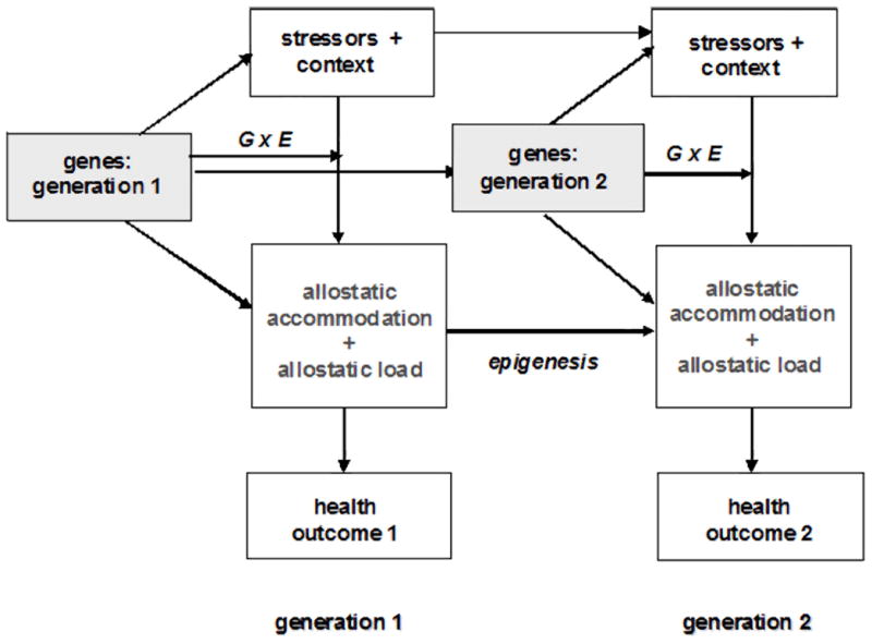
a. Schematic illustration of the midline frontal-brainstem axis involved in brain-body information transfer regulating peripheral autonomic, endocrine, and immune function. We argue that this axis is a key component of central allostatic accommodation, as discussed here (reprinted from Neuroimage, 47, R.D. Lane, & T.D. Wager, The new field of brain-body medicine: What have we learned and where are we headed? p. 1136 (2009), with permission from Elsevier). b. Figure showing areas of significantly lower gray matter volume (in yellow) in healthy adults who were within a mile and a half of the World Trade Center on 9/11/01 more than three years prior to imaging, relative to comparison group (reprinted from Neuroimage, 40, B. Ganzel, P. Kim, G. Glover, & E. Temple Resilience after 9/11: Multimodal neuroimaging evidence for stress-related change in the healthy adult brain. 791, (2008), with permission from Elsevier).
This overlap is at the heart of our allostatic model of the current stress process, in which the brain structures that evaluate the significance of emotional stimuli and produce emotional and behavioral response -- and act to drive regulatory responses and physiology peripheral to the CNS – are expected to be critically influenced by individual differences in central allostatic load. This is an emerging area of stress research; there are very few studies that link prior life stress, current brain function and/or structure, and current autonomic, endocrine, or immune function in healthy humans (although see Eisenberger et al., 2007; Ganzel, Kim et al., 2007). Yet from our theoretical model, we would predict that individual differences in central allostatic load influence all (Sterling, 2004) or almost all (McEwen, 2004) regulatory functions, and that this is a key predictor of long-term health and mortality. In short, the core emotional regions of the brain (including brainstem nuclei) will be the first to show evidence of short-term plasticity and long-term “wear-and tear” as a consequence of accumulating stressor exposure, and this will translate to short- or long-term alterations in autonomic, endocrine, and immune function, and peripheral physiology. This provides an organizing framework for associating social and physical context with mental and physical health.
Allostasis and cognition, in context
Stress-related disregulation of the amygdala and of the core emotional regions of the brain also has clear implications for ongoing emotional cognition. Based on our theoretical model, we would predict that there are long-term effects of accumulating environmental stressor exposure on emotion regulation (e.g., Ochsner et al., 2002; Kim & Hamann, 2004; Phan et al, 2004), emotional memory (Adolphs, Tranel, & Buchanan, 2005; Sharot, Martorella, Delgado, & Phelps, 2007), and control of attention to threat-related stimuli (e.g., Bishop, Duncan, Brett, & Lawrence, 2004) in healthy adults and children. The role of social context in this model suggests that these effects will be expected to be larger in low-SES environments in direct proportion of the the extent to which SES is associated with acute and chronic stressor exposure and diminution of protective resources. The addition of genetics into this model suggests that there will be cross-generational continuity in these effects, as well as important individual differences in vulernability and resilience to the acquisition of allostatic load.
Further, the core emotional systems of the brain are closely linked to the prefrontal neural systems responsible for executive function and general cognition (Pessoa, 2008), so that stress-related disruptions in one set of structures should drive disruptions in the other. For example, if allostatic load induces chronic hyperactivity of the amygdala, this is likely to have cascading effects on executive function in both children and adults, whether or not there is presence of stress-related clinical disorder. This is particularly true because the limbic system powerfully co-opts neural resources and attention (e.g., Anderson, 2005; Ohman, Flykt, & Esteves, 2001; Vuilleumier, Armony, Driver, & Dolan, 2001). Recent work on the development of executive function skills (Blair, 2002; Rothbart, Sheese, & Posner, 2007) has highlighted the links between the behavioral manifestations of emotion regulation, on the one hand (Blair, 2002; Rothbart et al., 2007), and the neural areas that underlie executive function, on the other (e.g., the anterior cingulate gyrus and lateral prefrontal areas; Fan, Flombaum, McCandliss, Thomas, & Posner, 2003; Fan, McCandliss, Fossella, Flombaum, & Posner, 2005). Notably, decreases in executive functioning have been observed in children living in lower socioeconomic status (SES) environments (Noble, Norman, & Farah, 2005). Children in lower SES environments have also been observed to have increased cortisol levels (Lupien, King, Meaney, & McEwen, 2000), which are an indicator of increased central allostatic load, as discussed in prior sections of this paper. We therefore predict that load-related alterations in the core emotional systems of the brain play a significant causal role in the observed decreases in executive function in low SES children (e.g., Blair, 2009).
Allostasis and mental disorder
Future research is also needed to clarify the contribution of stress and central allostatic load to the etiology and neural systems associated with mental disorder. The current understanding of the underlying neural mechanisms of each specific disorder is likely to be confounded with the more general effects of allostatic load on the brain. For example, amygdala gray matter atrophy has been reported in a number of mental disorders, including anxiety disorder (Milham et al., 2005), depression (Campbell et al., 2004; Siegle, Konecky, Thase, Carter, 2003; Sheline et al., 1998), borderline personality disorder (Schmahl et al., 2003) and autism (Dalton et al., 2007; Nacewicz et al., 2006; Schumann et al., 2004). Amygdala hyperexcitability has also been reported in these populations (Dalton et al., 2005; Siegle et al., 2003; Schmahl et al., 2003; Fales et al., 2008; Thomas et al., 2001). In depression, for example, the amygdala is consistently reported to be hyperexcitable (e.g., Fales et al., in press; Sheline et al., 2001). However, the amygdala is hypertrophic in early depression (Frodl et al., 2002) but shows atrophy after multiple episodes of major depression (Sheline et al., 1998). It has been argued (McEwen, 2003) that that chronic amygdala hyperexcitability in depression may itself produce these changes in amygdala volume over time, possibly through the actions of excitatory amino acids. In particular, the loss of glial cells, which protect surrounding tissue against neurotoxic damage, has been posited as a potential underlying mechanism for volume loss in amygdala and prefrontal cortex in depression (Sheline, 2003). This is also a plausible mechanism for the decreases in amygdala volume in healthy adults who were nearest the terrorist attacks of 9/11/01, since decreased amygdala volume was associated with increased amygdala hyperexcitability (Ganzel et al., 2008). As discussed above, amygdala hyperexcitability and reduced amygdala volume were also associated with higher overall levels of lifetime trauma exposure in that sample (Ganzel et al., 2008). Because trauma exposure is not uncommon in the general population (Kessler et al., 1995) and because mental illness is itself a stressor, we predict that stress plays a common explanatory role in the differences in amygdala structure and function observed across these mental disorders (i.e., that stress is a common factor explaining a significant portion of the variance in these outcomes). If so, this process would be characterized by equifinality, i.e., multiple paths to a single physiological outcome. This would be an important step in distinguishing those neural differences that do play a unique role in the etiology of each of these disorders. Examination of this question would require a convergence of expertise from multiple domains of stress research, including the social-psychological measurement of stressors and contextual risk, the clinical understanding of disorder, and neuroscience techniques. Careful assessment of stressor exposure in both clinical and control groups in neuroimaging studies of clinical disorder would shed light on this issue, as would longitudinal studies of amygdala excitability and volume over time in healthy trauma-exposed samples.
Allostasis and aging
As an additional point for future research, there is considerable overlap between the areas showing stress-related decreases in gray matter volume in humans (Ganzel et al., 2008; Gianaros, Jennings et al., 2007) and animals (e.g., Cerqueira et al., 2005) and the areas of greatest gray matter atrophy observed in normal aging (Resnick, Pham, Kraut, Zonderman, & Davatzikos, 2003; Shiino et al., 2006). This highlights the question of whether the gray matter volume differences observed in aging are better characterized as stress-related allostatic load, age-related atrophy, or both. Because the accumulation of stressor exposure experienced in ones’s lifetime will increase with age (for example, a longer lifespan provides increased opportunity for exposure to psychological trauma: e.g., Ganzel et al., 2008), we predict that lifetime stressor exposure is a significant contributor to the gray matter changes observed in normal aging. These effects are expected to be exacerbated within genetically stress-vulnerable individuals (gene by environment interactions: Figure 5). There is, to date, only limited information on the accumulating impact of life stress on the aging human brain. Here again, this question falls on the interface of stress research in the life sciences and the social and psychological sciences and would require a convergence of expertise to address.
Figure 5.

Linking health outcomes across generations: the role of genetics in a simplified allostatic model of stress includes the main effects of genetic inheritance on the stress physiology and choice of environment, gene × environment (G × E) interactions that affect the stress response, and epigenesis as a potential means to inherit parental allostatic load.
Moderators of allostasis
This allostatic model also suggests approaches for predicting important moderators of the stress process. If the brain is the central mediator of allostasis, the biological capacities and organization of the brain can help to determine the properties of a stressor that are likely to influence mental and physical health outcomes in the allostatic model. For example, a stressor must have sufficient magnitude to activate the emotional circuitry of the brain or the stress response will not be invoked by the organism; conversely, stressors that are of a magnitude sufficient to overwhelm the mechanisms of allostatic accommodation will produce greater allostatic load. A stressor’s duration and/or chronicity is also likely to modify the allostatic response; a stressor of sufficient persistence to exhaust (or render toxic) the biological processes of accommodation can produce profound effects on allostatic load, as previously discussed. We have also noted the expanding evidence that stressors of different types may be processed differently within the emotional circuitry of the brain, as, for example, with the neural processing of social pain versus physical threat (for reviews, see Alleva & Santucci, 2001; Dedovic, Duchesne, Andrews, Engert, & Pruessner, 2009; Depue & Morrone-Stupinsky, 2005; Eisenberger & Lieberman, 2004, Lieberman, 2007). Likewise, there is evidence that the developmental timing of stressor exposure can moderate central allostasis (e.g., Liu et al, 1997; Susser & Stein, 1994), We have reviewed evidence here that significant amounts of central and peripheral allostatic load may be acquired in adulthood; thus, when long-term biological effects of stressor exposure are observed in children, it remains to be determined what is different about the mechanisms of allostasis and allostatic load in early human development and how the long-term health consequences of allostatic load acquired in childhood may be distinct from the consequences of load acquired in adulthood. This special role of developmental timing in allostasis may derive from the development of knowledge of emotion (Tottenham, Leon, Casey, 2006), from differences in maturation time across the emotional circuitry of the brain (Casey, Getz, & Galvan, 2008), from a biological stress-sensitive period (analogous to the well-established stress sensitivity in the first ten days of a rat pup’s life: e.g., Levine, 2001; Liu et al., 2000), or a combination of these. It remains for future research to distinguish the impact of all of these potential moderators within the allostatic model of the current stress process.
Distinction between the differing impact of developmental timing and stressor type is of particular importance for studies of the consequences of early stressor exposure. The most frequently-studied stressors of childhood involve separation from (or conflict with) the primary attachment figure (e.g., parental loss, neglect, rejection, and/or conflict). These are stressors that fit the description of social pain. As previously discussed, there is evidence for qualitative physiological differences in the neural processing of physical threat relative to that of social pain, regardless of age at time of stressor exposure (e.g., Dedovic et al., 2009; Eisenberger & Lieberman, 2004). If the processing of social pain and physical threat are biologically distinguishable within the core emotional systems of the brain, then the allostatic model would predict that central and peripheral allostatic load accrues in distinguishable physiological systems for each type of stressor. This, in turn, should lead to different long-term health outcomes for each type of stressor (there is preliminary evidence for this, e.g., Post, Leverich, Xing, and Weiss, 2001). If stressor type indeed moderates the stress process in a way that is independent of developmental timing, it will be important to include stressor type as a separate variable in models of early stress. Furthermore, the effects of stressors of different types may change across the course of development, so that developmental timing and stressor type interact with once another (a three-way interaction).
Together, these considerations point to the need for attention to likely moderators of allostasis in studies of stress, as well as highlighting opportunites for new research. These may include, for example, further research into differences over time in the physiological processes underlying central and peripheral allostasis and allostatic load in response to stressors of different intensity, chronicity, type, and developmental timing, which in turn would be expected to drive multifinality in outcomes associated with long-term health and behavior. Longitudinal studies of amygdala responsiveness over time in samples of different ages and with different types and levels of stressor exposure would shed light on these points. Such work would also clarify the time frames for the development of stress-related alteration in brain structure and function, and their possible reversal. This is precisely the kind of work that can be built from the model we present.
Allostasis and the “tipping point”
A final consideration concerns the “tipping point” at which allostatic accommodation begins to generate maladaptive amounts of allostatic load, and thus begin to have long-term negative implications for physical and mental health. This is consistent with the notion of discontinuous change in dynamic systems theory (discussed earlier) and it raises the question of whether there is a neural and behavioral continuum from adaptive stress-related increases in vigilance and/or avoidance to the presence of a diagnosable clinical disorder. If so, this suggests multiple points for intervention to prevent accrual of allostatic load in nonclinical stress-exposed populations. If there is not such a continuum, or if it differs significantly among individuals, what are the unique factors that differentiate the “normal” stress process from those stress processes that predict pathology? Adoption of an allostatic model of the stress process provides impetus towards clarifying these important relationships and building a stronger understanding of the biology of resilience (Rutter, 1983a).
Conclusion
Exploiting the brain’s role as the central mediator of allostasis, we develop a model of the interplay over time between current stressor exposure (in its physical, social, and cultural context), the internal regulation of bodily processes, and health outcomes, giving us an integrated “bioecological model” (Bronfenbrenner & Morris, 1998) of the stress process. In support of this model, we have reviewed evidence that the core emotional systems of the brain serve together as the first and primary mediator of physiological accommodation to a current stressor (allostatic accommodation), and that all other stress-related physiological and behavioral change follows from this central mediation. Identification of the brain areas that are preferentially recruited to mediate allostasis highlights these regions as the most likely to show the physiological “wear and tear” due to allostatic load. This hypothesis is supported by recent human neuroimaging data; to date, the core emotional systems of the brain are observed to be the neural systems most likely to show long-term structural and functional differences following environmental challenge. We noted that these effects are observable both early and later in development in individuals who do not have a clinical disorder, suggesting that central allostatic accommodation and allostatic load are not pathological (although they may confer vulnerability to pathology), nor do they require a developmental sensitive period. This supports the general application of our model of the interplay over time between context, stressor exposure, internal regulation of bodily processes, and health outcomes. We also illustrated how this model facilitates the integration of current findings in human neuroscience and genetics with key elements of stress models from the social and life sciences, with implications for future research.
This model also has implications for the design of interventions targeting individuals at risk. A number of researchers have been on the cutting edge of this work in examining the biological mechanisms underlying the effectiveness of interventions designed to decrease the impact of prior stressors on health. Such studies are critical to the further development of theoretical models of the stress process. Intervention research provides explicit information about the potential plasticity of the organism—what is changeable in the individual and the means (intervention effort) by which is it possible to produce such change. Inclusion of measures of central and peripheral accommodation and central and peripheral indicators of load in intervention research would allow better assessment of the malleability of the biological systems implicated in the stress process and better ways to test concrete hypotheses about the drivers of these systems. However, there are only a handful of studies testing intervention effects on physiological outcomes and even fewer that examine the neural mediators of these processes. It is our hope that by setting forth a testable framework for linking stressors in the external environment with mental and physical health outcomes through stress-related plasticity in the brain, this allostatic model of the stress process will generate new studies that include measurement across the multiple levels of analysis included in our theoretical model.
There are a handful of notable examples of cross-cutting work that have tested environmental influences on physiological indicators of allostatic accommodation and load by including physiological measurement into randomized-control trials of interventions. For example, Gaab et al., (2003) assessed the effects of short-term cognitive-behavioral therapy on cortisol response to the Trier Social Stress Test and cognitive appraisal. In a small sample of male students, these researchers found convincing evidence for an effect of their short-term therapeutic intervention on cortisol response to the stress test. These effects were mediated by primary stress appraisal and self efficacy appraisal assessed via self report. Another interesting example of research linking psychosocial intervention and physiological measures of peripheral accommodation is Fisher and colleague’s work with foster care families (Philip, Fisher, Stoolmiller, Gunnar, & Burraston, 2007). In that work, a behavioral training intervention targeted at foster care parents of preschool children was found to lead to change in HPA axis functioning among the children by stabilizing levels of cortisol over the course of the day. More specifically, that work showed a more consistent pattern of cortisol change across the day in the treatment group children and less variability in evening cortisol levels, when compared to the randomized control group. In healthy adults, a randomized, controlled study of the effects of a brief (8-week) mindful-meditation stress reduction program has found significant increases in left-sided brain activation (associated with positive affect) which were correlated with increased antibody titers to influenza vaccine in the group receiving the intervention (Davidson et al., 2003). These effects may underlie the significant psychiatric symptom reduction and improvements in wound healing associated with similar mindful meditation interventions in clinical populations (e.g., Carmody & Baer, in press; Kabat-Zinn et al., 1992; 1998; Miller, Fletcher, & Kabat-Zinn, 1995). In terms of our proposed model, these studies make it clear that intervention is capable of having significant influences on both central and peripheral allostatic accommodation to current stressors. These, in turn, are likely to be playing a key causal role in the association between these interventions and their benefits.
Overall, these studies illustrate that intervention research is critically important in understanding the sensitivity of these stress-responsive systems to intervention. At the same time, studies that incorporate measures of the multiple components of the model presented here, including context, central and peripheral indicators of load, and mental and physical health outcomes, can further clarify the pathways by which stress gets “under the skin”. It is our expectation that measurement of the processes mediating the effect of intervention on physiogical mechanisms and behavioral outcomes will allow the design of more targeted interventions aimed at the primary drivers of systemic change.
A good example of research that would add substantially to the literature on stress and brain processes are exeperimental studies that target family resources. A number of studies launched in the late 1990s tested approaches to increasing income and the self sufficiency of low-income parents—some good examples are the New Hope evaluation (Huston et al., 2001), the Minnesota Family Investment Program (Gennetian & Miller, 2002), and the Canadian Self Sufficiency Project (Morris & Michalopoulos, 2003). Designed as randomized-control trials, one group of low-income parents in each of these studies was offered a substantial increase in their income (by allowing them to keep more of their welfare income or by supplementing their earned income) and another was only provided with the standard set of benefits available to low-income families. Notably, benefits to children were observed as a result of parents’ random assignment to the intervention group – with young children in the intervention groups having higher achievement in school (by about 15–20 percent of a standard deviation in magnitude) as compared to their peers assigned to the comparison group. Measures of achievement were based on teacher reports, parents reports, and direct assessments of children’s cognitive performance.
These studies have been used to causally link income and outcomes for children, using methodological techniques designed to isolate the income effects from other changes occurring as a result of the intervention with families (ie., increased employment effort; Morris, Duncan, & Rodrigues, 2006; Morris, Gennetian, & Duncan, 2005; Gennetian, Magnuson, & Morris, 2008). Unfortunately, these studies did not include assessments of either central or peripheral indicators of allostatic load. If they had, we could have learned a great deal about the neural and peripheral mechanisms relating increases in family resources and the resulting gains in children’s acheivement. Comparisons between poor and nonpoor children will get us only so far in understanding the stress process and its implications for the human brain; integrating longitudinal measures of central allostatic load into intervention studies will allow us to test models of plasticity and change in these neural processes much more precisely. To the extent that additional income to poor families provides protection from stressor exposure, such studies would be tests of the allostatic model presented here.
In setting the agenda for stress research nearly two decades ago, Rutter (1983a) called for research on individual differences in vulnerability and resilience in person-environment interactions and a better understanding of the interplay between stressor exposure and later outcomes in the context of development. This agenda has been updated to emphasize the need for an understanding of the moderating and mediating mechanisms underlying the association between risk and outcome, in order to create contextual models of risk and resilience (Cohen, Kessler, & Gordon, 1995; Gore and Eckenrode, 1996). All of these as-yet-unfulfilled agenda items are furthered by the adoption of a concrete allostatic model of the stress process that includes the influence of past and current stressors, environmental context, and the biology of the current stress response in the prediction of physical and mental health outcomes. Adoption of this model has the potential to expand the effectiveness of intervention for at-risk populations at the clinical and community level. Now is precisely the time for this work because of recent advances in stress research, including the emergence of allostasis as a theoretical platform and the development of new data collection methods for measuring multiple components of this theoretical model. With these tools at hand, researchers are now equipped to begin a new era in stress research that benefits from convergent perspectives in the biological and social sciences.
Acknowledgments
The authors wish to thank John Eckenrode, Valerie Reyna, Henry Ricciuti, Barbara Finlay, Steve Robertson, and Richard Depue for their helpful comments on earlier versions of the manuscript. The development of this work was supported in part by the National Institute of Mental Health (F31 MH63544, F32 MH068139, PI B.L. Ganzel) and the National Institute on Aging (P50 AG1711-06, project 3, PI Elaine Wethington; P30 AG022845, PI K. Pillemer), and the National Institute on Drug Abuse (U01 DA023821, PI T. Kamarck).
Footnotes
The core emotional regions are those areas of the brain that serve as hubs (Pessoa, 2008; Honey, Kotter, Breakspear, & Sporns, 2007) in the neural networks used to process affectively-laden information, including information regarding stressors.
There is debate in the field whether allostasis requires essentially all physiological parameters to co-vary with behavioral state (thus replacing homeostasis: Sterling & Eyer, 1988, Sterling, 2004) or whether a subset of systems retains homeostatic inflexibility (in which case allostasis updates homeostasis but does not replace it: e.g., McEwen, 2004; Wingfield, 2004). CNS control of most regulatory systems remains fundamental to both views.
The extended amygdala consists of several central forebrain structures with similar morphology and connectivity, including the bed nucleus of the stria terminalis, the central medial amygdala, the posterior shell of the nucleus accumbens, and the sublenticular substantia innominata (Alheid & Heimer, 1988).
However, this “privileged status” is vulnerable to high attentional load and highly competing sensory input (e.g., Pessoa, McKenna, Guitierrez, & Ungerlieder, 2002; Pessoa, Japee, Sturman, & Ungerleider, 2006).
This would be true even for systemic stressors (e.g., hemorrhage or infection), which are first registered viscerally and are unlikely to be under command or executive control of the emotional systems of the brain in the initial stages of processing (Anisman et al., 2003; Li, Ericsson, & Sawchenko, 1996; Mason, 1971; Pacak & Palkovits, 2001). However, Levine and Ursin (1991) have argued that even stressors that do not appear overtly “psychological” are likely to activate emotional and cognitive neural responses because they include novelty, expectation, contextual association and will arouse efforts towards avoidance because they are noxious.
There may, however, be important gender differences in behavioral response to threat that are mediated by oxytocin (“fight or flight” versus “tend and befriend”: Taylor et al., 2000).
The human dorsolateral PFC has been argued to be homologous with the rat medial PFC (Brown & Bowman, 2002).
Publisher's Disclaimer: The following manuscript is the final accepted manuscript. It has not been subjected to the final copyediting, fact-checking, and proofreading required for formal publication. It is not the definitive, publisher-authenticated version. The American Psychological Association and its Council of Editors disclaim any responsibility or liabilities for errors or omissions of this manuscript version, any version derived from this manuscript by NIH, or other third parties. The published version is available at www.apa.org/journals/rev
Contributor Information
Barbara L. Ganzel, Department of Human Development, Cornell University, Ithaca, NY
Pamela A. Morris, Department of Applied Psychology, Steinhardt School of Culture, Education, and Human Development, New York University, New York, NY
Elaine Wethington, Department of Human Development and Department of Sociology, Cornell University, Ithaca, NY.
References
- Adamec RE, Blundell J, Burton P. Neural circuit changes mediating lasting brain and behavioral response to predator stress. Neuroscience and Biobehavioral Review. 2005;29:1225–1241. doi: 10.1016/j.neubiorev.2005.05.007. [DOI] [PubMed] [Google Scholar]
- Adolphs R. Is the human amygdala specialized for processing social information? New York Academy of Sciences. 2003;985:319–353. doi: 10.1111/j.1749-6632.2003.tb07091.x. [DOI] [PubMed] [Google Scholar]
- Adolphs R, Tranel D, Buchanan TW. Amygdala damage impairs emotional memory for gist but not details of complex stimuli. Nature Neuroscience. 2005;8:512–518. doi: 10.1038/nn1413. [DOI] [PubMed] [Google Scholar]
- Albeck D, McKittrick C, Blanchard DC, Blanchard R, Nikulina J, McEwen B, Sakai R. Chronic social stress alters levels of corticotropin-releasing factor and arginine vasopressin mRNA in rat brain. The Journal of Neuroscience. 1997;17:4895–4903. doi: 10.1523/JNEUROSCI.17-12-04895.1997. [DOI] [PMC free article] [PubMed] [Google Scholar]
- Albert TJ, Dempesy CW, Sorenson CA. Anterior cerebellar vermal stimulation: effect on behavior and basal forebrain neurochemistry in rat. Biological Psychiatry. 1985;20:1267–1276. doi: 10.1016/0006-3223(85)90111-8. [DOI] [PubMed] [Google Scholar]
- Alleva E, Santucci D. Psychosocial vs. “physical” stress situations in rodents and humans. Physiology & Behavior. 2001;73:313–320. doi: 10.1016/s0031-9384(01)00498-x. [DOI] [PubMed] [Google Scholar]
- Altemus M, Gold PW. Neurohormones in depression and anxiety. In: Schulkin J, editor. Hormonally induced changes in the mind and brain. San Diego, CA: Academic Press; 1993. pp. 253–286. [Google Scholar]
- Amaral DG, Behniea H, Kelly JL. Topographic organization of the projections from the amygdala to the visual cortex of the macaque monkey. Neuroscience. 2003;118:1099–120. doi: 10.1016/s0306-4522(02)01001-1. [DOI] [PubMed] [Google Scholar]
- Ames EW. The development of Romanian orphanage children adopted to Canada. Canada: 1997. Final report to Human Resources Development. [Google Scholar]
- Anderson AK. Affective influences on the attentional dynamics supporting awareness. Journal of Experimental Psychology General. 2005;134:258–281. doi: 10.1037/0096-3445.134.2.258. [DOI] [PubMed] [Google Scholar]
- Anisman H, Merali Z, Hayley S. Sensitization associated with stressors and cytokine treatments. Brain, Behavior, and Immunity. 2003;17:86–93. doi: 10.1016/s0889-1591(02)00100-9. [DOI] [PubMed] [Google Scholar]
- Antonovsky A. Conceptual and methodological problems in the study of resistance resources and stressful life events. In: Dohrenwend BS, Dohrenwend BP, editors. Stressful life events: Their nature and effects. New York: John Wiley & Sons; 1974. pp. 245–257. [Google Scholar]
- Arborelius L, Owens M, Plotsky P, Nemeroff C. The role of corticotropin-releasing factor in depression and anxiety. Journal of Endocrinology. 1999;160:1–12. doi: 10.1677/joe.0.1600001. [DOI] [PubMed] [Google Scholar]
- Armony JL, Dolan RJ. Modulation of spatial attention by fear-conditioned stimuli: an event-related fMRI study. Neuropsychologia. 2002;40:817–26. doi: 10.1016/s0028-3932(01)00178-6. [DOI] [PubMed] [Google Scholar]
- Armony JL, LeDoux J. How the brain processes emotion. In: Yehuda R, McFarlane A, editors. Psychobiology of posttraumatic stress disorder. 821 . New York: The New York Academy of Sciences; 1997. pp. 259–271. [DOI] [PubMed] [Google Scholar]
- Backs RW. A comparison of factor analytic methods of obtaining cardiovascular autonomic components for the assessment of mental workload. Ergonomics. 1998;41(13):733–745. doi: 10.1080/001401398186883. [DOI] [PubMed] [Google Scholar]
- Baltes PB, Baltes MM. Psychological perspectives on successful aging: The model of selective optimization with compensation. In: Baltes PB, Baltes MM, editors. Successful aging: Perspectives from the behavioral sciences. New York: Cambridge University Press; 1990. pp. 1–34. [Google Scholar]
- Bandura A. Self-efficacy: Towards a unifying model of behavioral change. Psychological Review. 1977;84:191–215. doi: 10.1037//0033-295x.84.2.191. [DOI] [PubMed] [Google Scholar]
- Barton RA, Aggleton J. Primate evolution and the amygdala. In: Aggleton JP, editor. The Amygdala. 2. New York: Oxford University Press; 2000. pp. 479–508. [Google Scholar]
- Baum A, Garofalo J, Yali AM. Socioeconomic status and health in industrial nations: Social, psychological, and biological pathways. In: Adler NE, Marmot M, editors. Annals of the New York Academy of Sciences. 896 . New York: New York Academy of Sciences; 1999. pp. 131–144. [Google Scholar]
- Bechara A. A neural view of the regulation of complex cognitive functions by emotion. In: Philippot P, Feldman RS, editors. The Regulation of Emotion. Mahwah, NJ: Lawrence Erlbaum Associates; 2004. pp. 3–32. [Google Scholar]
- Bechara A, Damasio H, Damasio AR. Role of the amygdala in decision-making. New York Academy of Sciences. 2003;985:356–369. doi: 10.1111/j.1749-6632.2003.tb07094.x. [DOI] [PubMed] [Google Scholar]
- Bechara A, Damasio H, Damasio AR, Anderson S. Insensitivity to future consequences following damage to human prefrontal cortex. In: Mehler J, Franck S, editors. Cognition on Cognition. Cambridge, MA: MIT Press; 1995. pp. 3–12. [DOI] [PubMed] [Google Scholar]
- Bechara A, Tranel D, Damasio H, Adolps R, Rockland C, Damasio AR. Double dissociation of conditioning and declarative knowledge relative to the amaygdala and hippocampus in humans. Science. 1995;269:1115–1118. doi: 10.1126/science.7652558. [DOI] [PubMed] [Google Scholar]
- Becker GS. A Treatise on the Family. Cambridge, MA: Harvard University Press; 1981. [Google Scholar]
- Beers J. Orbitofrontal cortex and social regulation. In: Cacioppo JT, Visser PS, Pickett CL, editors. Social neuroscience: People thinking about people. Cambridge, MA: MIT Press; 2006. pp. 153–166. [Google Scholar]
- Belsky J. The determinants of parenting: A process model. Child Development. 1984;55:83–96. doi: 10.1111/j.1467-8624.1984.tb00275.x. [DOI] [PubMed] [Google Scholar]
- Bernard C. Lecons sur les phenomenones de la vie communs aux animaux et aux vegetaux. Paris: Ballieres; 1878. [Google Scholar]
- Berntson GG, Bechara A, Damasio A, Tranel D, Cacioppo JT. Amygdala contribution to selective dimensions of emotion. Social Cognitive Affective Neuroscience. 2007;2:123–129. doi: 10.1093/scan/nsm008. [DOI] [PMC free article] [PubMed] [Google Scholar]
- Bierhaus A, Wolf J, Andrassy M, Rohleder N, Humpert PM, Petrov D, Ferstl R, von Eynatten M, Wendt T, Rudofsky G, Joswig M, Morcos M, Schwaninger M, McEwen B, Kirschbaum C, Nawroth PP. A mechanism converting psychosocial stress into mononuclear cell activation. Proceedings of the National Academy of Sciences, USA. 2003;100:1920–1925. doi: 10.1073/pnas.0438019100. [DOI] [PMC free article] [PubMed] [Google Scholar]
- Bishop S, Duncan J, Brett Lawrence AD. Prefrontal cortical function and anxiety: controlling attention to threat-related stimuli. Nature Neuroscience. 2004;7:184–188. doi: 10.1038/nn1173. [DOI] [PubMed] [Google Scholar]
- Blair C. School readiness: Integrating cognition and emotion in a neurobiological Conceptualization of children’s functioning at school entry. American Psychologist. 2002;57:111–127. doi: 10.1037//0003-066x.57.2.111. [DOI] [PubMed] [Google Scholar]
- Blanchard R, McKittrick C, Blanchard D. Animal models of social stress: Effects on behavior and brain neurochemical systems. Physiology & Behavior. 2001;73:261–271. doi: 10.1016/s0031-9384(01)00449-8. [DOI] [PubMed] [Google Scholar]
- Blanchard DC, Sakai BS, McEwen BS, Weiss SM, Blanchard RJ. Subordinate stress: Behavioral, brain, and neuroendocrine correlates. Behavioral Brain Research. 1993;58:113–121. doi: 10.1016/0166-4328(93)90096-9. [DOI] [PubMed] [Google Scholar]
- Blanchard DC, Spencer R, Wiess S, Blanchard R, McEwen B, Sakai R. Visible burrow system as a model of chronic social stress: Behavioral and neuroendocrine correlates. Psychoneuroendocrinology. 1995;20(2):117–134. doi: 10.1016/0306-4530(94)e0045-b. [DOI] [PubMed] [Google Scholar]
- Bolger N. Coping as a personality process: A prospective study. Journal of Personality and Social Psychology. 1990;59:525–537. doi: 10.1037//0022-3514.59.3.525. [DOI] [PubMed] [Google Scholar]
- Boothroyd RA, Evans ME. Environmental safety and exposure to violence of inner city children experiencing a psychiatric crisis. International Journal of Anthropology. 2001;16:197–209. [Google Scholar]
- Bordi F, LeDoux J. Response properties of single units in areas of rat auditory thalamus that project to the amygdala, II. Cells receiving convergent auditory and somatosensory inputs and cells antidromatically activated by amygdala stimulation. Experimental Brain Research. 1994;98:275–286. doi: 10.1007/BF00228415. [DOI] [PubMed] [Google Scholar]
- Borokovia LV, Ivanova S, Zhang M, Yang H, Botchkina GI, Watkins LR, Wang H, Abumrad N, Eaton JW, Tracey KJ. Vagus nerve stimulation attentuates the systemic inflammatory response to endotoxin. Nature. 2000;405:458–462. doi: 10.1038/35013070. [DOI] [PubMed] [Google Scholar]
- Boulos Z, Rosenwasser AM. A chronobiologica perspective on allostasis and its application to shift work. In: Schulkin J, editor. Allostasis, homeostasis, and the costs of physiological adaptation. Cambridge, MA: Cambridge University Press; 2004. pp. 228–301. [Google Scholar]
- Bramsen I, Dirkzwager AJE, van der Ploeg HM. Predeployment personality traits and exposure to trauma as predictors of posttraumatic stress symptoms: A prospective study of form peacekeepers. American Journal of Psychiatry. 2000;157:1115–1119. doi: 10.1176/appi.ajp.157.7.1115. [DOI] [PubMed] [Google Scholar]
- Bremner JD, Licinio J, Darnell A, Krystal J, Owens M, Southwick S, Nemeroff C, Charney D. Elevated CSF corticotropin releasing factor concentrations in posttraumatic stress disorder. American Journal of Psychiatry. 1997;154:624–629. doi: 10.1176/ajp.154.5.624. [DOI] [PMC free article] [PubMed] [Google Scholar]
- Bremner JD, Randall P, Scott T, Bronen R, Southwick S, Seibyl J, Delaney R, McCarthy G, Charney D, Innis R. MRI-based measurement of hippocampal volume in patients with posttraumatic stress disorder. American Journal of Psychiatry. 1995;152:973–981. doi: 10.1176/ajp.152.7.973. [DOI] [PMC free article] [PubMed] [Google Scholar]
- Bremner JD, Vermetten E. Stress and development: Behavioral and biological consequences. Development and Psychopathology. 2001;13:473–490. doi: 10.1017/s0954579401003042. [DOI] [PubMed] [Google Scholar]
- Bremner JD, Southwick S, Johnson D, Yehuda R, Charney D. Childhood physical abuse and combat-related posttraumatic stress disorder in Vietnam veterans. American Journal of Psychiatry. 1993;150:235–239. doi: 10.1176/ajp.150.2.235. [DOI] [PubMed] [Google Scholar]
- Breslau N, Kessler RC, Chilcoat HD, Schultz LR, Davis GC, Andreski P. Trauma and posttraumatic stress disorder in the community - The 1996 Detroit Area Survey of Trauma. Archives of General Psychiatry. 1998;57 doi: 10.1001/archpsyc.55.7.626. [DOI] [PubMed] [Google Scholar]
- Broekman B, Olff M, Boer F. The genetic background of PTSD. Neuroscience and Biobehavioral Reviews. 2007;31:348–362. doi: 10.1016/j.neubiorev.2006.10.001. [DOI] [PubMed] [Google Scholar]
- Brody G. Prevention effects moderate the association of 5-HTTLPR and youth risk behavior initiation: GxE Hypotheses tested via a randomized prevention design; Presentation at the S. Kellam Festshrift, Johns Hopkins School of Public Health; 2008. [DOI] [PubMed] [Google Scholar]
- Bronfenbrenner U, Morris P. The ecology of developmental processes. In: Damon W, Lerner RM, editors. Theoretical Models of Human Development: Vol. 1. Handbook of Child Psychology. 5. New York: Wiley; 1998. pp. 993–1028. [Google Scholar]
- Bronfenbrenner U, Morris P. The Bioecological Model of Human Develoment. In: Lerner RM, Damon W, editors. Theoretical Models of Human Development. Vol. 1 Handbook of Child Psychology. 6. New York: Wiley; 2006. pp. 793–828. [Google Scholar]
- Brooks-Gunn J, Duncan G, Aber JL. Neighborhood Poverty. New York: Russell Sage Foundation; 1997. [Google Scholar]
- Brown GW. Life events and affective disorder: Replications and limitations. Psychosomatic Medicine. 1993;55:248–259. doi: 10.1097/00006842-199305000-00003. [DOI] [PubMed] [Google Scholar]
- Brown GW. Social roles, context, and evolution in the origin of depression. Journal of Health and Social Behavior. 2002;43:255–276. [PubMed] [Google Scholar]
- Brown GW, Harris TO. ocial origins of depression: A study of depressive disorder in women. New York: Free Press; 1978. S. [Google Scholar]
- Brown GW, Harris TO. Life events and illness. New York and London: The Guildford Press; 1989. [Google Scholar]
- Brown VJ, Bowman EM. Rodent models of prefrontal cortex function. Trends in Neuroscience. 2002;25:340–343. doi: 10.1016/s0166-2236(02)02164-1. [DOI] [PubMed] [Google Scholar]
- Brunner EJ, Hemingway H, Walker BR, Page, MClarke P, Juneja M. Adrenocortical, Autonomic, and Inflammatory Causes of the Metabolic Syndrome: Nested Case-Control Study. Circulation. 2002;106:2659–2665. doi: 10.1161/01.cir.0000038364.26310.bd. [DOI] [PubMed] [Google Scholar]
- Buchel C, Morris J, Dolan RJ, Friston KJ. Brain systems mediating aversive conditioning: an event-related fMRI study. Neuron. 1998;20:947–957. doi: 10.1016/s0896-6273(00)80476-6. [DOI] [PubMed] [Google Scholar]
- Burchfield S, Woods S, Elich M. Pituitary adreno-cortical response to chronic intermittant stressors. Physiology and Behavior. 1980;24:297–302. doi: 10.1016/0031-9384(80)90090-6. [DOI] [PubMed] [Google Scholar]
- Bush G, Luu P, Posner MI. Cognitive and emotional influences in anterior cingulate cortex. Trends in Cognitive Science. 2000;4:215–22. doi: 10.1016/s1364-6613(00)01483-2. [DOI] [PubMed] [Google Scholar]
- Cacioppo JT, Amaral DG, Blanchard JJ, Cameron JL, Carter CS, Crews D, Fiske S, Heatherton T, Johnson MK, Kozak MJ, Levenson RW, Lord C, Miller EK, Ochsner K, Raichle ME, Shea MT, Taylor SE, Young LJ, Quinn KJ. Social neuroscience: Progress and implications for mental health. Perspectives on Psychological Science. 2007;2:99–123. doi: 10.1111/j.1745-6916.2007.00032.x. [DOI] [PubMed] [Google Scholar]
- Cacioppo JT, Amaral DG, Blanchard JJ, Cameron JL, Carter CS, Crews D, Fiske S, Heatherton T, Johnson MK, Kozak MJ, Levenson RW, Lord C, Miller EK, Ochsner K, Raichle ME, Shea MT, Taylor SE, Young LJ, Quinn KJ. Social neuroscience: Progress and implications for mental health. Perspectives on Psychological Science. 2007;2:99–123. doi: 10.1111/j.1745-6916.2007.00032.x. [DOI] [PubMed] [Google Scholar]
- Cacioppo JT, Berntson GG. Social psychological contributions to the decade of the brain. American Psychologist. 1992;47(8):1019–1028. doi: 10.1037//0003-066x.47.8.1019. [DOI] [PubMed] [Google Scholar]
- Cacioppo JT, Berntson GG. Relationship between attitudes and evaluative space: A critical review, with emphasis on the separability of positive and negative substrates. Psychological Bulletin. 1994;115:401–423. [Google Scholar]
- Cacioppo JT, Berntson GG. The brain, homeostasis, and health: Balancing demands of the internal and external milieu. In: Friedman HS, Cohen Silver R, editors. Foundations of Health Psychology. New York: Oxford University Press; 2007. pp. 73–91. [Google Scholar]
- Cacioppo JT, Berntson GG, Sheridan JF, McClintock MK. Multilevel integrative analyses of human behavior: Social neuroscience and the complementing nature of social and biological approaches. Psychological Bulletin. 2000;126:829–843. doi: 10.1037/0033-2909.126.6.829. [DOI] [PubMed] [Google Scholar]
- Cacioppo J, Ernst J, Burleson M, McClintock M, Malarkey W, Hawkley L, Kowalewski RA, Hobson JA, Hugdahl K, Spiegel D, Berntson G. Lonely traits and concomitant physiological processes: The MacArthur Social Neuroscience Studies. International Journal of Psychophysiology. 2000;35:143–154. doi: 10.1016/s0167-8760(99)00049-5. [DOI] [PubMed] [Google Scholar]
- Cacioppo JT, Gardner WL. Emotion. Annual Review of Psychology. 1999;50:191–214. doi: 10.1146/annurev.psych.50.1.191. [DOI] [PubMed] [Google Scholar]
- Campbell S, Marriott M, Nahmias C, MacQueen GM. Lower hippocampal volume in patients suffering from depression: A meta-analysis. American Journal of Psychiatry. 2004;161:598–607. doi: 10.1176/appi.ajp.161.4.598. [DOI] [PubMed] [Google Scholar]
- Canli T, Qui M, Omura K, Congdon E, Haas BW, Amin Z, Herrmann MJ, Constable RT, Lesch KP. Neural Correlates of Epigenesis. Proceedings of the National Academy of Sciences U S A. 2006;103:16033–16038. doi: 10.1073/pnas.0601674103. [DOI] [PMC free article] [PubMed] [Google Scholar]
- Cannon WB. Bodily changes in pain, hunger, fear and rage: An account of recent searches into the function of emotional excitement. New York: D. Appleton and Co; 1920. [Google Scholar]
- Cannon WB. The wisdom of the body. New York: W. W. Norton; 1932. [Google Scholar]
- Cannon WB. Stresses and strains of homeostasis. American Journal of Medical Science. 1935;189:1–14. [Google Scholar]
- Carmody J, Baer R. Relationships between Mindfulness Practice and Levels of Mindfulness, Medical and Psychological Symptoms and Well-being in a Mindfulness-Based Stress Reduction Program. Journal of Behavioral Medicine. doi: 10.1007/s10865-007-9130-7. in press. [DOI] [PubMed] [Google Scholar]
- Casey BJ, Getz S, Galvan A. The adolescent brain. Developmental Review. 2008;28:62–77. doi: 10.1016/j.dr.2007.08.003. [DOI] [PMC free article] [PubMed] [Google Scholar]
- Casey BJ, Tottenham N, Fossella J. Clinical, imaging, lesion, and genetic approaches toward a model of cognitive control. Developmental Psychobiology. 2002;40:237–254. doi: 10.1002/dev.10030. [DOI] [PubMed] [Google Scholar]
- Casey BJ, Glatt CE, Tottenham N, Soliman F, Bath K, Amso D, et al. BDNF as a model system for examining gene x environment interactions across development. Neuroscience. doi: 10.1016/j.neuroscience.2009.03.081. in press. [DOI] [PMC free article] [PubMed] [Google Scholar]
- Caspi A, McClay J, Moffitt T, Mill J, Martin J, Craig I, Taylor A, Poulton R. Role of geneotype in cycle of violence in maltreated children. Science. 2002;297:851–853. doi: 10.1126/science.1072290. [DOI] [PubMed] [Google Scholar]
- Caspi A, Sugden K, Moffitt TE, Taylor A, Craig I, Harrington H, McClay J, Mill J, Martin J, Braithwaite A, Poulton R. Influence of life stress on depression: Moderation by a polymorphism in the 5-HTT gene. Science. 2003;301:386–389. doi: 10.1126/science.1083968. [DOI] [PubMed] [Google Scholar]
- Cerqueira JJ, Pego JM, Taipa R, Bessa JM, Almeida OFX, Sousa N. Morphological correlates of corticosteroid-induced changes in prefrontal cortex-dependent behaviors. The Journal of Neuroscience. 2005;25:7792–7800. doi: 10.1523/JNEUROSCI.1598-05.2005. [DOI] [PMC free article] [PubMed] [Google Scholar]
- Cerqueira JJ, Mailliet F, Almeida OFX, Jay TM, Sousa N. The prefrontal cortex as a key target of the maladaptive response to stress. The Journal of Neuroscience. 2007;27:2781–2787. doi: 10.1523/JNEUROSCI.4372-06.2007. [DOI] [PMC free article] [PubMed] [Google Scholar]
- Charney D, Grillon C, Bremner JD. The neurobiological basis of anxiety and fear: Circuits, mechanisms, and neurochemical interactions (part I) The Neuroscientist. 1998;4(1):35–44. [Google Scholar]
- Chrousos G, Gold P. The concepts of stress and stress system disorders: Overview of physical and behavioral homeostasis. Journal of the American Medical Association. 1992;267:1244–1252. [PubMed] [Google Scholar]
- Cicchetti D, Rogosch F. Diverse patterns of neuroendocrine activity in maltreated children. Psychopathology and Development. 2001;13:677–694. doi: 10.1017/s0954579401003145. [DOI] [PubMed] [Google Scholar]
- Coan JA, Schaefer HS, Davidson RJ. Lending a hand: social regulation of the neural response to threat. Psychological Science. 2006;17:1032–1039. doi: 10.1111/j.1467-9280.2006.01832.x. [DOI] [PubMed] [Google Scholar]
- Cobb S. Social support as a moderator of life stress. Psychosomatic Medicine. 1976;38:300–314. doi: 10.1097/00006842-197609000-00003. [DOI] [PubMed] [Google Scholar]
- Cohen RA, Grieve S, Hoth KF, Paul RH, Sweet L, Tate D, Gunstad J, Stroud L, McCaffery J, Hitsman B, Niaura R, Clark CR, McFarlane A, Bryant R, Gordon E, Williams LM. Early life stress and morphometry of the adult anterior cingulate cortex and caudate nuclei. Biological Psychiatry. 2006;59:975–982. doi: 10.1016/j.biopsych.2005.12.016. [DOI] [PubMed] [Google Scholar]
- Cohen S, Doyle WJ, Turner RB, Alper C, Skoner DP. Emotional style and susceptibility to the common cold. Psychosomatic Medicine. 2002;65:652–657. doi: 10.1097/01.psy.0000077508.57784.da. [DOI] [PubMed] [Google Scholar]
- Cohen S, Kessler RC, Gordon LU. Strategies for measuring stress in studies of pyschiatric and physical disorders. In: Cohen S, Kessler RC, Gordon LU, editors. Measuring stress: A guide for health and social scientists. New York: Oxford University Press; 1995. pp. 3–28. [Google Scholar]
- Cohen S, Tyrrell D, Smith A. Psychological stress and susceptibility to the common cold. New England Journal of Medicine. 1991;325:606–612. doi: 10.1056/NEJM199108293250903. [DOI] [PubMed] [Google Scholar]
- Colcombe SJ, Kramer AF, Erickson KI, Scalf P, McAuley E, Cohen NJ, et al. Cardiovascular fitness, cortical plasticity, and aging. Proceedings of the National Academy of Sciences. 2003;101(9):3316–3321. doi: 10.1073/pnas.0400266101. [DOI] [PMC free article] [PubMed] [Google Scholar]
- Cole SW, Hawkley LC, Arevalo JM, Sung CY, Rose RM, Cacioppo JT. Social regulation of gene expression in human leukocytes. Genome Biology. 2007;8:R189.1–R189.13. doi: 10.1186/gb-2007-8-9-r189. [DOI] [PMC free article] [PubMed] [Google Scholar]
- Coleman JS. Social capital in the creation of human capital. American Journal of Sociology. 1988;94:S95–S120. [Google Scholar]
- Convit A, Wolf OT, Tarshish C, de Leon MJ. Reduced glucose tolerance is associated with poor memory performance and hippocampal atrophy among normal elderly. Proceedings of the National Academy of Sciences USA. 2003;100:2019–2022. doi: 10.1073/pnas.0336073100. [DOI] [PMC free article] [PubMed] [Google Scholar]
- Coplan J, Andrews M, Rosenblum L, Owens M, Friedman S, Gorman J, Nemeroff C. Persistent elevations of cerebrospinal fluid concentrations of corticotropin-releasing factor in adult non-human primates exposed to early-life stressors: Implications for the pathophysiology of mood and anxiety disorders. Proceedings of the National Academy of Sciences USA. 1996;93:1619–1623. doi: 10.1073/pnas.93.4.1619. [DOI] [PMC free article] [PubMed] [Google Scholar]
- Coplan J, Trost R, Owens M, Cooper T, Gorman J, Nemeroff C, Rosenblum L. Cerebrospinal fluid concentrations of somatostatin and biogenic amines in grown primates reared by mothers exposed to manipulated foraging conditions. Archives of General Psychiatry. 1998;55:473–477. doi: 10.1001/archpsyc.55.5.473. [DOI] [PubMed] [Google Scholar]
- Dahl G, Lochner L. The impact of family income on child achievement: Evidence from the Earned Income Tax Credit. Cambridge, MA: National Bureau of Economic Research; 2008. NBER Working Paper Series: Working Paper 14599. [Google Scholar]
- Dalton KM, Nacewicz BM, Alexander AL, Davidson RJ. Gaze-fixation, brain activation, and amygdala volume in unaffected siblings of individuals with autism. Biological Psychiatry. 2007;61:512–20. doi: 10.1016/j.biopsych.2006.05.019. [DOI] [PubMed] [Google Scholar]
- Dalton KM, Nacewicz BM, Johnstone T, Schaefer HS, Gernsbacher MA, Goldsmith HH, et al. Gaze fixation and the neural circuitry of face processing in autism. Nature Neuroscience. 2005;8:519–26. doi: 10.1038/nn1421. [DOI] [PMC free article] [PubMed] [Google Scholar]
- Davidson RJ. Affective neuroscience: The case for interdisciplinary research. In: Kessel F, Rosenfeld P, Anderson N, editors. Expanding the boundaries of health and social sciences. Oxford: Oxford University Press; 2003. pp. 99–121. [Google Scholar]
- Davidson R, Jackson D, Kalin N. Emotion, plasticity, context, and regulation: Perspectives from affective neuroscience. Psychological Bulletin. 2000;126:890–908. doi: 10.1037/0033-2909.126.6.890. [DOI] [PubMed] [Google Scholar]
- Davidson R, Maxwell JS, Shackman AJ. Privileged status of emotion in the brain. Proceedings of the National Academy of Sciences U S A. 2004;101:11919–11916. doi: 10.1073/pnas.0404264101. [DOI] [PMC free article] [PubMed] [Google Scholar]
- Davidson RJ, Kabat-Zinn J, Schumacher J, Rosenkranz M, Muller D, Santorelli SF, et al. Alterations in brain and immune function produced by mindfulness meditation. Psychosomatic Medicine. 2003;65:564–570. doi: 10.1097/01.psy.0000077505.67574.e3. [DOI] [PubMed] [Google Scholar]
- Davis M. The Role of the Amygdala in Fear and Anxiety. Annual Review of Neuroscience. 1999;15:353–375. doi: 10.1146/annurev.ne.15.030192.002033. [DOI] [PubMed] [Google Scholar]
- Davis M, Walker D, Lee Y. Roles of the amygdala and bed nucleus of the stria terminalis in fear and anxiety measured with the acoustic startle reflex: Possible relevance to PTSD. In: Yehuda R, McFarlane A, editors. Psychobiology of posttraumatic stress disorder. 821 . New York: The New York Academy of Sciences; 1997. pp. 305–331. [DOI] [PubMed] [Google Scholar]
- Dedovic K, Duchesne A, Andrews J, Engert V, Pruessner JC. The brain and the stress axis: The neural correlates of cortisol regulation in response to stress. NeuroImage. 2009;47:864–871. doi: 10.1016/j.neuroimage.2009.05.074. [DOI] [PubMed] [Google Scholar]
- Depue R, Morrone-Strupinsky J. A neurobiological model of affiliative bonding: Implications for conceptualizing a human trait of affiliation. Behavioral and Brain Science. 2005;28:313–350. doi: 10.1017/S0140525X05000063. [DOI] [PubMed] [Google Scholar]
- Dickerson SS, Kemeny ME. Acute stressors and cortisol responses: A theoretical integration and synthesis of laboratory research. Psychological Bulletin. 2004;130:355–391. doi: 10.1037/0033-2909.130.3.355. [DOI] [PubMed] [Google Scholar]
- Dodic M, Moritz K, Wintour E. Prenatal exposure to glucocorticoids and adult disease. Archives of Physiology and Biochemistry. 2003;111:61–9. doi: 10.1076/apab.111.1.61.15144. [DOI] [PubMed] [Google Scholar]
- Dohrenwend BP. Theoretical integration. In: Dohrenwend BP, editor. Adversity, stress, and psychopathology. New York: Oxford University Press; 1998. pp. 523–555. [Google Scholar]
- Dohrenwend BP. Inventorying stressful life events as risk factors for psychopathology: Towards resolution of the problem of intracategory variability. Psycholgoical Bulletin. 2006;132:477–495. doi: 10.1037/0033-2909.132.3.477. [DOI] [PMC free article] [PubMed] [Google Scholar]
- Dohrenwend BS, Ashkenasy AR, Krasnoff L, Dohrenwend BP. Exemplification of a method for scaling life events: The PERI Life Events Scale. Journal of Health and Social Behavior. 1978;19:205–229. [PubMed] [Google Scholar]
- Dolan RJ, Vuilleumier P. Amygdala automaticity in emotional processing. Annals of the New York Academy of Sciences. 2003;985:348–355. doi: 10.1111/j.1749-6632.2003.tb07093.x. [DOI] [PubMed] [Google Scholar]
- Dolcos F, LaBar KS, Cabeza R. Dissociable effects of arousal and valence on prefrontal activity indexing emotional evaluation and subsequent memory: An event-related fMRI study. NeuroImage. 2004;23:64–74. doi: 10.1016/j.neuroimage.2004.05.015. [DOI] [PubMed] [Google Scholar]
- Drevets WC, Videen TO, Price JL, Preskorn SH, Carmichael ST, Raichle ME. A functional anatomical study of unipolar depression. Journal of Neuroscience. 1992;12:3628–3641. doi: 10.1523/JNEUROSCI.12-09-03628.1992. [DOI] [PMC free article] [PubMed] [Google Scholar]
- Drury SS, Theall KP, Keats BJB, Sheeringa M. The role of the dopamine transporter (DAT) gene in the development of PTSD in preschool children. Journal of Traumatic Stress. doi: 10.1002/jts.20475. in press. [DOI] [PMC free article] [PubMed] [Google Scholar]
- Duman RS, Monteggia LM. A neurotrophic model for stress-related mood disorders. Biological Psychiatry. 2006;59:1116–1127. doi: 10.1016/j.biopsych.2006.02.013. [DOI] [PubMed] [Google Scholar]
- Duncan GJ, Brooks-Gunn J, editors. Consequences of growing up poor. New York: Sage; 1997. [Google Scholar]
- Duncan GJ, Brooks-Gunn J. Family Poverty, Welfare Reform and Child Development. Child Development. 2000;71:188–196. doi: 10.1111/1467-8624.00133. [DOI] [PubMed] [Google Scholar]
- Duncan GJ, Kalil A, Ziol-Guest K. The economic costs of early childhood Poverty. 4. Washington, DC: Partnership for America‘s Economic Success; 2008. [Google Scholar]
- Duncan G, Morris P, Rodrigues C. Does money really matter? Estimating impacts of family income on children’s achievement with data from social policy experiments. New York: MDRC; 2006. Unpublished manuscript. [DOI] [PMC free article] [PubMed] [Google Scholar]
- Duncan GJ, Yeung WJ, Brooks-Gunn J, Smith JR. Does Poverty Affect the Life Chances of Children? American Sociological Review. 1998;63:406–23. [Google Scholar]
- Duncan G, Morris P, Rodrigues C. Estimating impacts of family income on children’s achievement with data from social policy experiments. New York: MDRC; 2006. Does money really matter? Unpublished Manuscript. [DOI] [PMC free article] [PubMed] [Google Scholar]
- Earls F, Buka S. Measurement of community characteristics. In: Meisels SJ, Shonkoff JP, editors. Handbook of Early Childhood Intervention. Cambridge, MA: Cambridge University Press; 2000. pp. 309–326. [Google Scholar]
- Eaton W. The sociology of mental disorders. New York: Greenwood Press; 2000. [Google Scholar]
- Eisenberger NI, Lieberman MD, Williams KD. Does rejection hurt? An fMRI study of social exclusion. Science. 2003;302:290–292. doi: 10.1126/science.1089134. [DOI] [PubMed] [Google Scholar]
- Eisenberger NI, Lieberman MD. Why rejection hurts: a common neural alarm system for physical and social pain. Trends in Cognitive Sciences. 2004;8:294–300. doi: 10.1016/j.tics.2004.05.010. [DOI] [PubMed] [Google Scholar]
- Eisenberger NI, Taylor SE, Gable SL, Hilmert CJ, Lieberman MD. Neural pathways link social support to attenuated neuroendocrine stress responses. NeuroImage. 2007;35:1601–1612. doi: 10.1016/j.neuroimage.2007.01.038. [DOI] [PMC free article] [PubMed] [Google Scholar]
- Evans GW. A multimethodological analysis of cumulative risk and allostatic load among rural children. Developmental Psychology. 2003;39:924–933. doi: 10.1037/0012-1649.39.5.924. [DOI] [PubMed] [Google Scholar]
- Evans GW. Child development and the physical environment. Annual Review of Psychology. 2006;57:423–451. doi: 10.1146/annurev.psych.57.102904.190057. [DOI] [PubMed] [Google Scholar]
- Evans GW, Kim P, Ting AH, Tesher HB, Shannis D. Cumulative risk, maternal responsiveness, and allostatic load among young adolescents. Developmental Psychology. 2007;43:341–35. doi: 10.1037/0012-1649.43.2.341. [DOI] [PubMed] [Google Scholar]
- Evans GW, Saltzman H, Cooperman JL. Housing Quality and Children’s Socioemotional Health. Environment and Behavior. 2001;33(3):389–399. [Google Scholar]
- Fales CL, Barch DM, Rundle MM, Mintun MA, Snyder AZ, Cohen JD, Mathews J, Sheline YI. Altered emotional interference processing in affective and cognitive-control brain circuitry in major depression. Biological Psychiatry. 2008;63:377–84. doi: 10.1016/j.biopsych.2007.06.012. [DOI] [PMC free article] [PubMed] [Google Scholar]
- Fan J, Flombaum JI, McCandliss BD, Thomas KM, Posner MI. Cognitive and brain consequences of conflict. Neuroimage. 2003;18:42–57. doi: 10.1006/nimg.2002.1319. [DOI] [PubMed] [Google Scholar]
- Fan J, McCandliss BD, Fossella J, Flombaum JI, Posner MI. The activation of attentional networks. Neuroimage. 2005;26:471–479. doi: 10.1016/j.neuroimage.2005.02.004. [DOI] [PubMed] [Google Scholar]
- Farah MJ, Betancourt L, Shera DM, Savage JH, Giannetta JM, Brodsky NL, et al. Environmental stimulation, parental nuturance and cognitive development in humans. Developmental Science. doi: 10.1111/j.1467-7687.2008.00688.x. in press. [DOI] [PubMed] [Google Scholar]
- Farah MJ, Shera DM, Savage JH, Betancourt L, Giannetta JM, Brodsky NL, et al. Childhood poverty: specific associations with neurocognitive development. Brain Research. 2006;111:166–174. doi: 10.1016/j.brainres.2006.06.072. [DOI] [PubMed] [Google Scholar]
- Feldman S, Conforti N, Itsik A, Weidenfeld J. The role in limbic structures in the modulation of ACTH responses following adrenalectomy. In: Chrousos G, McCarty R, Pacak K, Cizza G, Sternberg E, Gold P, Kvetnansky R, editors. Stress: Basic mechanisms and clinical implications. 771 . New York: The New York Academy of Sciences; 1995. pp. 73–91. [DOI] [PubMed] [Google Scholar]
- Philip A, Fisher P, Stoolmiller M, Gunnar M, Burrastona B. Effects of a therapeutic intervention for foster preschoolers on diurnal cortisol activity. Psychoneuroendocrinology. 2007;32:892–905. doi: 10.1016/j.psyneuen.2007.06.008. [DOI] [PMC free article] [PubMed] [Google Scholar]
- Foa E, Kozak M. Emotional processing of fear: Exposure to corrective information. Psychological Bulletin. 1986;99:20–35. [PubMed] [Google Scholar]
- Folkow B, Neil E. Circulation. London: Oxford University Press; 1971. [Google Scholar]
- Ford DH, Lerner RM. Developmental systems theory: An integrative approach. Thousand Oaks, CA: Sage; 1992. [Google Scholar]
- Forsyth R. Blood pressure responses to long-term avoidance schedules in the unrestrained rhesus monkey. Psychosomatic Medicine. 1969;31:300. doi: 10.1097/00006842-196907000-00003. [DOI] [PubMed] [Google Scholar]
- Francis DD, Meaney MJ. Maternal care and the development of stress responses. Current Opinion in Neurobiology. 1999;9:128–134. doi: 10.1016/s0959-4388(99)80016-6. [DOI] [PubMed] [Google Scholar]
- Frankenhaeuser M. Psychobiological aspects of life stress. In: Levine S, Ursin J, editors. Coping and Health. New York: Plenum Press; 1979. pp. 203–224. [Google Scholar]
- Freese JL, Amaral DG. The organization of projections from the amgydala to the visual cortical areas TE and V1 in the Macaque monkey. Journal of Comparative Neurology. 2005;486:295–317. doi: 10.1002/cne.20520. [DOI] [PubMed] [Google Scholar]
- Freitas A, Downey G. Resilience: A dynamic perspective. International Journal of Behavior Development. 1998;22:263–285. [Google Scholar]
- Frodl T, Meisenzahl E, Zetzsche T, Bottlender R, Born C, Groll C, et al. Enlargement of the amygdala in patients with a first episode of major depression. Biological Psychiatry. 2002;51:708–714. doi: 10.1016/s0006-3223(01)01359-2. [DOI] [PubMed] [Google Scholar]
- Funayama ES, Grillon C, Davis M, Phelps EA. A double dissociation in the affective modulation of startle in humans: Effects of unilateral temporal lobectomy. Journal of Cognitive Neuroscience. 2001;13:721–29. doi: 10.1162/08989290152541395. [DOI] [PubMed] [Google Scholar]
- Furstenberg FF, Jr, Brooks-Gunn J, Morgan SP. Adolescent mothers in later life. Cambridge, MA: Cambridge University Press; 1987. [PubMed] [Google Scholar]
- Gaab J, Blattler N, Menzi T, Pabst B, Stoyer S, Ehlert U. Randomized controlled evaluation of the effects of cognitive-behavioral stress management on cortisol responses to acute stress in healthy subjects. Psychoneuroendocrinology. 2003;28:767–779. doi: 10.1016/s0306-4530(02)00069-0. [DOI] [PubMed] [Google Scholar]
- Gallo L, Matthews K. Understanding the association between socioeconomic status and physical health: Do negative emotions play a role? Psychological Bulletin. 2003;129:10–51. doi: 10.1037/0033-2909.129.1.10. [DOI] [PubMed] [Google Scholar]
- Galvan A, Hare TA, Parra CE, Penn J, Voss H, Glover G, Casey BJ. Earlier development of the accumbens relative to orbitofrontal cortex might underlie risk-taking behavior in adolescents. Journal of Neuroscience. 2006;26:2885–2892. doi: 10.1523/JNEUROSCI.1062-06.2006. [DOI] [PMC free article] [PubMed] [Google Scholar]
- Ganzel B, Casey BJ, Glover G, Voss H, Temple E. The aftermath of September 11th: Effect of intensity and recency of trauma on outcome. Emotion. 2007;7:227–238. doi: 10.1037/1528-3542.7.2.227. [DOI] [PMC free article] [PubMed] [Google Scholar]
- Ganzel B, Kim P, Altemus M, Voss HU, Temple E. After 9/11: Grey matter density in right ventromedial prefrontal cortex (vmPFC) predicts salivary cortisol levels in healthy trauma- exposed adults. Annals of the New York Academy of Sciences. 2007;1121:664–665. [Google Scholar]
- Ganzel B, Kim P, Glover G, Temple E. Resilience after 9/11: Multimodal neuroimaging evidence for stress-related change in the healthy adult brain. NeuroImage. 2008;40:788–795. doi: 10.1016/j.neuroimage.2007.12.010. [DOI] [PMC free article] [PubMed] [Google Scholar]
- Garmezy N. Reflections and commentary on risk, resilience, and development. In: Haggerty RJ, Sherrod LR, Garmezy N, Rutter M, editors. Stress, risk, and resilience in children and adolescents: Processes, mechanisms, and interventions. New York: Cambridge University Press; 1996. pp. 1–18. [Google Scholar]
- Gennetian LA, Miller C. Children and welfare reform: A view from an experimental welfare program in Minnesota. Child Development. 2002;73(2):601–620. doi: 10.1111/1467-8624.00426. [DOI] [PubMed] [Google Scholar]
- Gennetian L, Magnuson K, Morris P. From statistical association to causation: What developmentalists can learn from Instrumental Variables techniques coupled with experimental data. Developmental Psychology. 2008;44(2):381–394. doi: 10.1037/0012-1649.44.2.381. [alphabetic listing of authors to reflect equal contribution] [DOI] [PMC free article] [PubMed] [Google Scholar]
- Gerfen CR. The Neostriatal Mosaic: Multiple levels of compartmental organization in the basal ganglia. Annual Review of Neuroscience. 1992;15:285–320. doi: 10.1146/annurev.ne.15.030192.001441. [DOI] [PubMed] [Google Scholar]
- Geronimus AT, Hicken M, Keene D, Bound J. “Weathering” and age patterns of allostatic load scores among Blacks and Whites. American Journal of Public Health. 2006;96:826–833. doi: 10.2105/AJPH.2004.060749. [DOI] [PMC free article] [PubMed] [Google Scholar]
- Gershoff ET. Low income and the development of America’s kindergartners. New York: National Center for Children in Poverty; 2003. Living at the Edge Research Brief 4. [Google Scholar]
- Gershoff ET, Aber JL, Raver CC. Child poverty in the United States: An evidence-based framework for programs and policies. In: Lerner RM, Jacobs F, Wertlieb D, editors. Promoting positive child, adolescent and family development: A handbook of program and policy innovations. Thousand Oaks, CA: Sage Publications; 2003. pp. 81–136. [Google Scholar]
- Gianaros PJ, Horenstein JA, Cohen S, Matthews KA, Brown SM, et al. Perigenual anterior cingulate morphology covaries with perceived social standing. Social Cognition and Affective Neuroscience. 2007;2:161–73. doi: 10.1093/scan/nsm013. [DOI] [PMC free article] [PubMed] [Google Scholar]
- Gianaros PJ, Jennings JR, Sheu LK, Greer PJ, Kuller LH, Matthews KA. Prospective reports of chronic life stress predict decreased grey matter volume in the hippocampus. NeuroImage. 2007;35:795–803. doi: 10.1016/j.neuroimage.2006.10.045. [DOI] [PMC free article] [PubMed] [Google Scholar]
- Gianaros PJ, Horenstein JA, Hariri AR, Sheu LK, Manuck SB, Matthews KA, Cohen S. Potential neural embedding of parental social standing. Social Cognitive and Affective Neuroscience. 2008;3:91–96. doi: 10.1093/scan/nsn003. [DOI] [PMC free article] [PubMed] [Google Scholar]
- Gianaros PJ, Sheu LK. A review of neuroimaging studies of stressor-evoked blood pressure reactivity: Emerging evidence for a brain-body pathway to coronary heart disease risk. Neuroimage. 2009;47:922–936. doi: 10.1016/j.neuroimage.2009.04.073. [DOI] [PMC free article] [PubMed] [Google Scholar]
- Glantz M, Sloboda Z. Analysis and reconceptualization of resilience. In: Glantz M, Johnson J, editors. Resilience and development: Positive life adaptations. New York: Kluwer/Plenum; 1999. pp. 109–128. [Google Scholar]
- Glascher J, Adolphs R. Processing of the arousal of subliminal and supraliminal emotional stimuli by the human amygdala. Journal of Neuroscience. 2003;23:10274–82. doi: 10.1523/JNEUROSCI.23-32-10274.2003. [DOI] [PMC free article] [PubMed] [Google Scholar]
- Glaser R, Kiecolt-Glaser JK, Malarkey WB, Sheridan JF. The influence of psychological stress on the immune response to vaccines. Annals of the New York Academy of Sciences. 1998;840:649–655. doi: 10.1111/j.1749-6632.1998.tb09603.x. [DOI] [PubMed] [Google Scholar]
- Glaser R, Pearson GR, Bonneau RH, Esterling BA, Atkinson C, Kiecolt-Glaser JK. Stress and the memory t-cell response to the Epstein-Barr virus in healthy medical students. Health Psychology. 1993;12:435–442. doi: 10.1037//0278-6133.12.6.435. [DOI] [PubMed] [Google Scholar]
- Gold SM, Dziobek I, Sweat V, Tirsi A, Rogers K, Bruehl H, Tsui W, Richardson S, Javier E, Convit A. Hippocampal damage and memory impairments as possible early brain complications of type 2 diabetes. Diabetologia. 2007;50:711–719. doi: 10.1007/s00125-007-0602-7. [DOI] [PubMed] [Google Scholar]
- Goldstein DS. Stress as a scientific idea: Homeostatic theory of stress and distress. Homeostasis. 1995a;36:117–215. [Google Scholar]
- Goldstein DS. Stress, Catecholamines, and Cardiovascular Disease. New York: Oxford University Press; 1995b. [Google Scholar]
- Gore S, Eckenrode JE. Context and process in research on risk and resilience. In: Haggerty RJ, Sherrod LR, Garmezy N, Rutter M, editors. Stress, risk, and resilience in children and adolescents: Processes, mechanisms, and interventions. Cambridge, MA: Cambridge University Press; 1996. pp. 19–63. [Google Scholar]
- Gottfried J, O’Doherty J, Dolan RJ. Encoding predictive reward value in human amygdala and orbitofrontal cortex. Science. 2003;301:1104–7. doi: 10.1126/science.1087919. [DOI] [PubMed] [Google Scholar]
- Gottlieb G. Normally occurring environmental and behavioral influences on gene activity: From central dogma to probabilistic epigenesis. Psychological Review. 1998;105:792–802. doi: 10.1037/0033-295x.105.4.792-802. [DOI] [PubMed] [Google Scholar]
- Gould E, McEwen BS, Tanapat P, Galea LA, Fuchs E. Neurogenesis in the dentate gyrus of the adult tree shrew is regulated by psychosocial stress and NMDA receptor activation. Journal of Neuroscience. 1997;17:2492–8. doi: 10.1523/JNEUROSCI.17-07-02492.1997. [DOI] [PMC free article] [PubMed] [Google Scholar]
- Gould E, Tanapat P, Rydel T, Hastings N. Regulation of hippocampal neurogenesis in adulthood. Biological Psychiatry. 2000;48:715–720. doi: 10.1016/s0006-3223(00)01021-0. [DOI] [PubMed] [Google Scholar]
- Graham Y, Heim C, Goodman S, Miller A, Nemeroff C. The effects of neonatal stress on brain development: Implications for psychopathology. Development and Psychopathology. 1999;11:545–565. doi: 10.1017/s0954579499002205. [DOI] [PubMed] [Google Scholar]
- Granger D, Hood K, Dreschel N, Sergeant E, Likos A. Developmental effects of early immune stress on aggressive, socially reactive, and inhibited behaviors. Development and Psychopathology. 2001;13:599–610. doi: 10.1017/s0954579401003108. [DOI] [PubMed] [Google Scholar]
- Gray TS. Functional and neuroanatomical relationships among the amygdala, basal forebrain, ventral striatum and cortex: an integrative discussion. Annals of the New York Academy of Science. 1999;877:439–444. doi: 10.1111/j.1749-6632.1999.tb09281.x. [DOI] [PubMed] [Google Scholar]
- Gregg T, Siegal A. Brain structures and neurotransmitters regulating aggression in cats: Implications for human aggression. Progress in Neuro-Psychopharmacology and Biolgical Psychiatry. 2001;25:91–140. doi: 10.1016/s0278-5846(00)00150-0. [DOI] [PubMed] [Google Scholar]
- Gross JJ. Emotion regulation: Affective, cognitive, and social consequences. Psychophysiology. 2002;39:281–291. doi: 10.1017/s0048577201393198. [DOI] [PubMed] [Google Scholar]
- Gunnar MR, Quevedo K. The neurobiology of stress and development. In: Fiske S, editor. Annual Review of Psychology. Vol. 58. 2007. pp. 145–174. [DOI] [PubMed] [Google Scholar]
- Gunnar M, Vasquez D. Low cortisol and a flattening of expected daytime rhythm: Potential indices of risk in human development. Development and Psychopathology. 2001;13:515–538. doi: 10.1017/s0954579401003066. [DOI] [PubMed] [Google Scholar]
- Gurvits T, Shenton M, Hokama H, Ohta H, Lasko N, Gilbertson M, Orr S, Kikinis R, Jolesz R, McCarley R, Pitman R. Magnetic resonance imaging study of hippocampal volume in chronic, combat-related posttraumatic stress disorder. Biological Psychiatry. 1996;40:1091–1099. doi: 10.1016/S0006-3223(96)00229-6. [DOI] [PMC free article] [PubMed] [Google Scholar]
- Haber SN, Kim KS, Mailly P, Calzavara R. Reward-related cortical inputs define a large striatal region in primates that interface with associative cortical connections, providing a substrate for incentive-based learning. Journal of Neuroscience. 2006;26:8368–8376. doi: 10.1523/JNEUROSCI.0271-06.2006. [DOI] [PMC free article] [PubMed] [Google Scholar]
- Hamer D. Rethinking behavior genetics. Science. 2002;298:71–72. doi: 10.1126/science.1077582. [DOI] [PubMed] [Google Scholar]
- Harenski CL, Hamann S. Neural correlates of regulating negative emotions related to moral violations. Neuroimage. 2006;30:313–24. doi: 10.1016/j.neuroimage.2005.09.034. [DOI] [PubMed] [Google Scholar]
- Hariri AR, Drabant EM, Munoz KE, Kolachana BS, Mattay VS, Egan MF, Weinberger DR. A susceptibility gene for affective disorders and the response of the human amygdala. Archives of General Psychiatry. 2005;62:146–152. doi: 10.1001/archpsyc.62.2.146. [DOI] [PubMed] [Google Scholar]
- Hariri AR, Mattay VS, Tessitore A, Kolachana BS, Fera F, Goldman D, Egan MF, Weinberger DR. Serotonin transporter genetic variation and the response of the human amygdala. Science. 2002;297:400–403. doi: 10.1126/science.1071829. [DOI] [PubMed] [Google Scholar]
- Hariri AR, Bookheimer SY, Mazziotta JC. Modulating emotional responses: Effects of a neocortical network on the limbic system. NeuroReport. 2000;11:43–48. doi: 10.1097/00001756-200001170-00009. [DOI] [PubMed] [Google Scholar]
- Harvell CD, Tollrian R. Why inducible defenses? In: Trollrian R, Harvell CD, editors. The Ecology and Evolution of Inducible Defenses. Princeton University Press; Princeton: 1999. pp. 3–9. [Google Scholar]
- Haveman RH, Wolfe BS. On the effects of investments in children. New York: Russell Sage Foundation; 1994. [Google Scholar]
- Hawkins N, Davies R, Holmes T. Evidence of pyschosocial factors in the development of pulmonary tuberculosis. American Review of Tuberculosis and Pulmonary Diseases. 1957;75:768–780. doi: 10.1164/artpd.1957.75.5.768. [DOI] [PubMed] [Google Scholar]
- Hawkley LC, Berntson GG, Engeland CG, Marucha PT, Mash CM, Cacioppo JT. Stress, aging, and resilience: Can accrued wear and tear be slowed? Canadian Psychology. 2005;46:115–125. [Google Scholar]
- Heim C, Nemeroff C. The impact of early adverse experiences on brain systems involved in the pathophysiology of anxiety and affective disorders. Biological Psychiatry. 1999;46:1509–1522. doi: 10.1016/s0006-3223(99)00224-3. [DOI] [PubMed] [Google Scholar]
- Heim C, Newport J, Heit S, Graham Y, Wilcox M, Bonsali R, Miller A, Nemeroff C. Pituitary-adrenal and autonomic responses to stress in women after sexual and physical abuse in childhood. Journal of the American Medical Association. 2000;284:592–597. doi: 10.1001/jama.284.5.592. [DOI] [PubMed] [Google Scholar]
- Heim C, Owens M, Plotsky P, Nemeroff C. Endocrine factors in the pathophysiology of mental disorders: Persistent changes in corticotropin releasing factor systems due to early life stress. Psychopharmacology Bulletin. 1997;33:185–192. [PubMed] [Google Scholar]
- Heinrichs S, Mensaghi F, Pich E, Britton K, Koob G. The role of CRF in behavioral aspects of stress. In: Chrousos G, McCarty R, Pacak K, Cizza G, Sternberg E, Gold P, Kvetnansky R, editors. Stress: Basic mechanisms and clinical implications. 771 . New York: The New York Academy of Sciences; 1995. pp. 92–113. [DOI] [PubMed] [Google Scholar]
- Hobfoll SE. Conservation of resources: A new attempt at conceptualizing stress. American Psychologist. 1989;44:513–524. doi: 10.1037//0003-066x.44.3.513. [DOI] [PubMed] [Google Scholar]
- Hobfoll S. Influence of culture, community, and the nested self in the stress process: Advancing conservation of resources theory. Applied Psychology An International Review. 2001;50:337–421. [Google Scholar]
- Hobfoll SE, Hall BJ, Canetti-Nisim D, Galea S, Johnson RJ, Palmieri PA. Refining our understanding of traumatic growth in the face of terrorism: Moving from meaning cognitions to doing what is meaningful. Applied Psychology. 2007;56:345–366. [Google Scholar]
- Hobfoll SE, Johnson RJ, Ennis N, Jackson AP. Resource loss, resource gain, and emotional outcomes among inner city women. Journal of Personality and Social Psychology. 2003;84:632–643. [PubMed] [Google Scholar]
- Holahan CJ, Moos RH, Holahan CK, Cronkite RC. Resource loss, resource gain, and depressive symptoms: A 10-year longitudinal model. Journal of Personality and Social Psychology. 1999;77:620–629. doi: 10.1037//0022-3514.77.3.620. [DOI] [PubMed] [Google Scholar]
- Holahan CJ, Moos RH, Holahan CK, Cronkite RC. Long-term posttreatment functioning among patients with unipolar depression: An integrative model. Journal of Consulting and Clinical Psychology. 2000;68:226–232. doi: 10.1037//0022-006x.68.2.226. [DOI] [PubMed] [Google Scholar]
- Holmes T, Rahe R. The social readjustment rating scale. Journal of Psychosomatic Research. 1967;11:213–218. doi: 10.1016/0022-3999(67)90010-4. [DOI] [PubMed] [Google Scholar]
- Honey CJ, Kotter R, Breakspear M, Sporns O. Network structure of cerebral cortex shaps functional connectivity on multiple time scales. Proceedings of the National Academy of Sciences. 2007;104:10240–10245. doi: 10.1073/pnas.0701519104. [DOI] [PMC free article] [PubMed] [Google Scholar]
- House JS, Kessler RC, Herzog AR, Mero R, Kinney A, Breslow M. Social stratification, age, and health. In: Schaie KW, Blazer D, House JS, editors. Aging, health behaviors, and health outcomes ( Hillsdale, NJ: Lawrence Erlbaum Associates; 1992. pp. 1–31. [Google Scholar]
- House J, Landis KR, Umberson D. Social relationships and health, Science. 1988;41:540–545. doi: 10.1126/science.3399889. [DOI] [PubMed] [Google Scholar]
- Hubbs-Tate L, Nation J, Krebs NF, Bellinger DC. Neurotoxicants, micronutrients, and social environments: Individual and combined effects on children’s development. Psychological Science in the Public Interest. 2006;6:57–121. doi: 10.1111/j.1529-1006.2005.00024.x. [DOI] [PubMed] [Google Scholar]
- Hugdahl K, Ohman A. Effects of instruction acquisition and extinction of electrodermal responses to fear-relevant stimuli. Journal of Experimental Psychology: Human Learning and Memory. 1977;3:608–618. [PubMed] [Google Scholar]
- Huston A, Duncan G, Granger R, Bos H, McLoyd V, Mistry R, Crosby D, Gibson C, Magnuson K, Romich J, Ventura A. Work-based anti-poverty programs for parents can enhance the school performance and social behavior of children. Child Development. 2001;72(1):318–337. doi: 10.1111/1467-8624.00281. [DOI] [PubMed] [Google Scholar]
- Ironson G, Antoni M, Schneiderman N, LaPerriere A, Klimas N, Lutgendorf S, Fletcher M. Stress management and psychogenic predictors of disease course in HIV-1 infections. In: Goodkin K, Visser A, editors. Psychoneuroimmunology: Stress, mental disorders, and health. Washington, DC: American Psychiatric Press; 2000. pp. 317–356. [Google Scholar]
- Ishai A, Pessoa L, Bickle C, Ungerleider L. Repetition suppression of faces is modulated by emotion. Proceedings of the National Academy of Sciences. 2004;101:9827–9832. doi: 10.1073/pnas.0403559101. [DOI] [PMC free article] [PubMed] [Google Scholar]
- Kabat-Zinn J, Massion AO, Kristeller J, Peterson LG, Fletcher K, Pbert L, Linderking W, Santorelli SF. Effectiveness of a meditation-based stress reduction program in the treatment of anxiety disorders. American Journal of Psychiatry. 1992;149:936–943. doi: 10.1176/ajp.149.7.936. [DOI] [PubMed] [Google Scholar]
- Kabat-Zinn J, Wheeler E, Light T, Skillings A, Scharf MS, Cropley TG, Hosmer D, Bernhard J. Influence of a mindfulness-based stress reduction intervention on rates of skin clearing in patients with moderate to severe psoriasis undergoing phototherapy (UVB) and photochemotherapy (PUVA) Psychosomatic Medicine. 1998;60:625–632. doi: 10.1097/00006842-199809000-00020. [DOI] [PubMed] [Google Scholar]
- Kannan Y. Neuroendocrine-immune network in stress. In: Hedrick H, editor. The Laboratory Mouse. New York: Academic Press; 2004. pp. 301–309. [Google Scholar]
- Kaplan H. Towards an understanding of resilience: A critical review of definitions and models. In: Glantz M, Johnson J, editors. Resilience and development: Positive life adaptations. New York: Kluwer/Plenum; 1999. pp. 17–84. [Google Scholar]
- Kaplan JR, Adams MR, Anthony MS, Morgan TM, Manuck SB, Clarkson TB. Dominant social status and contraceptive hormone treatment inhibit atherogenesis in premenopausal monkeys. Arteriosclerosis, thrombosis, and vascular biology. 1995;15:2094–2100. doi: 10.1161/01.atv.15.12.2094. [DOI] [PubMed] [Google Scholar]
- Kaplan JR, Manuck SB, Clarkson TB, Lusso FM, Taub DM. Social status, environment and atherosclerosis in cynomologus monkeys. Arteriosclerosis. 1982;2:359–368. doi: 10.1161/01.atv.2.5.359. [DOI] [PubMed] [Google Scholar]
- Kaplan JR, Manuck SB, Clarkson TB, Lusso FM, Taub DM, Miller EW. Social stress and atherosclerosis in normocholesterolemic cynomologus monkeys. Science. 1983;220:733–735. doi: 10.1126/science.6836311. [DOI] [PubMed] [Google Scholar]
- Kapp BS, Whalen PJ, Supple WF, Pascoe JP. Amygdaloid contributions to conditioned fear. In: Aggelton JP, editor. The amygdala: Neurobiological aspects of emotion, Memory, and Mental Dysfunction. New York: Wiley-Liss; 1992. pp. 229–254. [Google Scholar]
- Karlamangala AS, Singer BH, McEwen BS, Rowe JW, Seeman TE. Allostatic load as a predictor of functional decline: MacArthur studies of successful aging. Journal of Clinical Epidemiology. 2002;55:696–710. doi: 10.1016/s0895-4356(02)00399-2. [DOI] [PubMed] [Google Scholar]
- Kaufman J, Charney D. Effects of early stress on brain structure and function: Implications for understanding the relationship between child maltreatment and depression. Development and Psychopathology. 2001;13:451–142. doi: 10.1017/s0954579401003030. [DOI] [PubMed] [Google Scholar]
- Kempermann G, Kuhn HG, Gage FH. Experience-induced neurogenesis in the senescent dentate gyrus. Journal of Neuroscience. 1998;18:3206–3212. doi: 10.1523/JNEUROSCI.18-09-03206.1998. [DOI] [PMC free article] [PubMed] [Google Scholar]
- Kendler K, Gardner C. Monozgotic twins discordant for major depression: A preliminary exploration of the role of environmental experiences in the aetiology and course of illness. Pyschological Medicine. 2001;31:411–423. doi: 10.1017/s0033291701003622. [DOI] [PubMed] [Google Scholar]
- Kendler K, Gardner C, Neale M, Prescott A. Genetic risk for major depression in men and women: Similar or different heritabilities and same or partly distinct genes? Psychological Medicine. 2001;31:605–616. doi: 10.1017/s0033291701003907. [DOI] [PubMed] [Google Scholar]
- Kessler R, Sonnega A, Bromet E, Hughes M, Nelson C. Posttraumatic stress disorder in the National Comorbidity Study. Archives of General Psychiatry. 1995;52:1048–1059. doi: 10.1001/archpsyc.1995.03950240066012. [DOI] [PubMed] [Google Scholar]
- Kim SH, Hamann S. Neural correlates of positive and negative emotion regulation. Journal of Cognitive Neuroscience. 2004;19:776–798. doi: 10.1162/jocn.2007.19.5.776. [DOI] [PubMed] [Google Scholar]
- Kim H, Somerville LH, Johnstone T, Polis S, Alexander AL, Shin LM, Whalen PJ. Contextual modulation of amygdala responsivity to surprised faces. Journal of Cognitive Neuroscience. 2004;16:1730–45. doi: 10.1162/0898929042947865. [DOI] [PubMed] [Google Scholar]
- Kishiyama MM, Boyce WT, Jimenez AM, Perry LM, Knight RT. Socioeconomic disparities affect prefrontal function in children. Journal of Cognitive Neuroscience. 2009 doi: 10.1162/jocn.2009.21101. [DOI] [PubMed] [Google Scholar]
- Koob GF. Alcoholism: Allostasis and beyond. Alcoholism Clinical and Experimental Research. 2003;27:232–43. doi: 10.1097/01.ALC.0000057122.36127.C2. [DOI] [PubMed] [Google Scholar]
- Krieger N. Theories for social epidemiology in the 21st century: an ecosocial perspective. International Journal of Epidemiology. 2001;30:668–677. doi: 10.1093/ije/30.4.668. [DOI] [PubMed] [Google Scholar]
- LaBar KS, Gatenby JC, Gore JC, LeDoux JE, Phelps EA. Human amygdala activation during conditioned fear acquisition and extinction: A mixed-trial fMRI study. Neuron. 1998;20:937–45. doi: 10.1016/s0896-6273(00)80475-4. [DOI] [PubMed] [Google Scholar]
- Lane RD, Fink GR, Chau PM, Dolan RJ. Neural activation during selective attention to subjective emotional responses. NeuroReport. 1997;8:3969–3972. doi: 10.1097/00001756-199712220-00024. [DOI] [PubMed] [Google Scholar]
- Lane RD, Waldstein SR, Chesney MA, Jennings JR, Lovallo WR, Kozel PJ, et al. The rebirth of neuroscience in psychosomatic medicine, Part I: Historical context, methods, and relevant basic science. Psychosomatic Medicine. 2009;71:117–134. doi: 10.1097/PSY.0b013e31819783be. [DOI] [PubMed] [Google Scholar]
- Lane RD, Wager TD. The new field of brain-body medicine: What have we learned and where are we headed? Neuroimage. 2009;47:1135–1140. doi: 10.1016/j.neuroimage.2009.06.013. [DOI] [PubMed] [Google Scholar]
- Lange CG, James W. The Emotions. Baltimore: Williams & Wilkins; 1922. [Google Scholar]
- Law K, Stroud L, LaGasse L, Niaura R, Liu J, Lester B. Smoking during pregnancy and newborn neurobehavior. Pediatrics. 2003;111:1318–23. doi: 10.1542/peds.111.6.1318. [DOI] [PubMed] [Google Scholar]
- Lazarus R. Psychological stress and the coping process. New York: McGraw-Hill; 1966. [Google Scholar]
- Lazarus R. Emotion and adaptation. New York: Oxford University Press; 1991. [Google Scholar]
- Lazarus R. Coping theory and research: Past, present, and future. Psychosomatic Medicine. 1993a;55:234–247. doi: 10.1097/00006842-199305000-00002. [DOI] [PubMed] [Google Scholar]
- Lazarus R. From psychological stress to the emotions: A history of changing outlooks. Annual Revew of Psychology. 1993b;44:1–21. doi: 10.1146/annurev.ps.44.020193.000245. [DOI] [PubMed] [Google Scholar]
- Lazarus R. Stress and Emotion. New York: Springer; 1999. [Google Scholar]
- Lazarus R, Folkman S. Stress, appraisal, and coping. New York: Springer; 1984. [Google Scholar]
- LeDoux JE. Emotional memory systems in the brain. Behavioral Brain Research. 1993;58:69–79. doi: 10.1016/0166-4328(93)90091-4. [DOI] [PubMed] [Google Scholar]
- LeDoux J. Emotion: Clues from the brain. Annual Review of Psychology. 1995;46:209–235. doi: 10.1146/annurev.ps.46.020195.001233. [DOI] [PubMed] [Google Scholar]
- LeDoux J. The emotional brain. New York: Simon and Schuster; 1996. [Google Scholar]
- LeDoux J, Cicchetti P, Xagoraris A, Romanski L. The lateral amygdaloid nucleus: Sensory interface in the amygdala in fear conditioning. Journal of Neuroscience. 1990;10:1062–1069. doi: 10.1523/JNEUROSCI.10-04-01062.1990. [DOI] [PMC free article] [PubMed] [Google Scholar]
- Lee VE, Burkam DT. Inequality at the starting gate: Social background differences in achievement as children begin school. Washington, DC: Economic Policy Institute; 2002. [Google Scholar]
- Lesch KP. Gene-environment interaction and the genetics of depression. Journal of Psychiatry & Neuroscience. 2004;29:174–184. [PMC free article] [PubMed] [Google Scholar]
- Levine S. Primary social relationships influence the development of the hypothalamic-pituitary- adrenal axis in the rat. Physiology & Behavior. 2001;73:255–260. doi: 10.1016/s0031-9384(01)00496-6. [DOI] [PubMed] [Google Scholar]
- Levine S, Ursin H. What is stress? In: Brown MR, Koob GF, Rivier C, editors. Stress: Neurobiology and neuroendocrinology. New York: Marcel Dekker; 1991. pp. 3–22. [Google Scholar]
- Li HY, Ericsson A, Sawchenko PE. Distinct mechanisms underlie activation of hypothalamic neurosecretory neurons and their medullary catecholamine afferents in categorically different stress paradigms. Proceedings of the National Academy of Sciences. 1996;93:2359–2364. doi: 10.1073/pnas.93.6.2359. [DOI] [PMC free article] [PubMed] [Google Scholar]
- Lieberman MD. Social cognitive neuroscience: A review of core processes. Annual Reviews of Psychology. 2007;58:259–289. doi: 10.1146/annurev.psych.58.110405.085654. [DOI] [PubMed] [Google Scholar]
- Liston C, McEwen BS, Casey BJ. Psychosocial stress reversibly disrupts prefrontal processing and attentional control. Proceedings of the National Academy of Sciences. 2009;106:912–917. doi: 10.1073/pnas.0807041106. [DOI] [PMC free article] [PubMed] [Google Scholar]
- Liu D, Dioro D, Tannebaum B, Caldji C, Francis D, Freedman A, Sharma S, Pearson D, Plotsky PM, Meany MJ. Maternal care, hippocampal glucocorticoid receptors, and hypothalamic-pituitary-adrenal responses to stress. Science. 1997;277:1659–1662. doi: 10.1126/science.277.5332.1659. [DOI] [PubMed] [Google Scholar]
- Liu D, Dioro D, Day JC, Francis D, Meany MJ. Maternal care, hippocampal synaptogenesis and cognitive development in rats. Nature Neuroscience. 2000;3:799–806. doi: 10.1038/77702. [DOI] [PubMed] [Google Scholar]
- Lund-Johanson P. Hemodynamic concepts in essential hypertension. Triangle. 1984;23:12–24. [Google Scholar]
- Lupien SJ, King S, Meaney MJ, McEwen B. Child’s stress hormone levels correlate with mother’s socioeconomic status and depressive state. Biological Psychiatry. 2000;48:976–980. doi: 10.1016/s0006-3223(00)00965-3. [DOI] [PubMed] [Google Scholar]
- Lupien SJ, McEwen B. The acute effects of corticosteroids on cognition: integration of animal and human model studies. Brain Research Reviews. 1997;24:1–27. doi: 10.1016/s0165-0173(97)00004-0. [DOI] [PubMed] [Google Scholar]
- Magnusson D. Individual development: A holistic, integrated model. In: Moen P, Elder G, Luescher K, editors. Examining lives in context: Perspectives on the ecology of human development. Washington, DC: American Psychological Association; 1995. pp. 19–60. [Google Scholar]
- Maier SF, Watkins LR. Cytokines for psychologists: Implications of bidirectional immune-to-brain communication for understanding behavior, mood, and cognition. Psychological Review. 1998;105:83–107. doi: 10.1037/0033-295x.105.1.83. [DOI] [PubMed] [Google Scholar]
- Manuck SB, Marsland AL, Kaplan JR, Williams JK. The pathogenicity of behavior and its neuroendocrine mediation: An example from coronary heart disease. Psychosomatic Medicine. 1995;57:275–283. doi: 10.1097/00006842-199505000-00009. [DOI] [PubMed] [Google Scholar]
- Margarinos AM, McEwen BS. Stress-induced atrophy of apical dendrites of hippocampus CA3c neurons: involvement of glucocorticoid secretion and excitatory amino acid receptors. Neuroscience. 1995;69:89–98. doi: 10.1016/0306-4522(95)00259-l. [DOI] [PubMed] [Google Scholar]
- Magarinos AM, McEwen BS. Experimental diabetes in rats causes hippocampal dendritic and synaptic reorganization and increased glucocorticoid reactivity to stress. Procedings of the National Academy of Sciences USA. 2000;97(20):11056–11061. doi: 10.1073/pnas.97.20.11056. [DOI] [PMC free article] [PubMed] [Google Scholar]
- Marmot M. Social differentials in health within and between populations. Daedalus. 1994;123:197–216. [Google Scholar]
- Marmot MG, Kogevinas M, Elston MA. Social/economic status and disease. Annual Review of Public Health. 1987;8:111–135. doi: 10.1146/annurev.pu.08.050187.000551. [DOI] [PubMed] [Google Scholar]
- Marmot MG, Shipley MJ, Rose G. Inequalities in death - Specific explanations of a general pattern? The Lancet. 1984;1:1003–1006. doi: 10.1016/s0140-6736(84)92337-7. [DOI] [PubMed] [Google Scholar]
- Mason JW. A re-evaluation of the concept of ‘non-specificity’ in stress theory. Journal of Psychiatry Research. 1971;8:323–333. doi: 10.1016/0022-3956(71)90028-8. [DOI] [PubMed] [Google Scholar]
- Mason JW. Emotions as reflected in patterns of endocrine regulation. In: Levi L, editor. Emotions: Their parameters and measurement. New York: Raven Press; 1975a. pp. 143–181. [Google Scholar]
- Mason JW. A historical view of the stress field. Journal of Human Stress. 1975b;1:6–12. doi: 10.1080/0097840X.1975.9940399. [DOI] [PubMed] [Google Scholar]
- Masten AS, Garmezy N, Tellengen A, Pellegrini DS, Larkin K, Larson A. Competence and stress in school children: The moderating effects of individual and family qualities. Journal of Child Psychology and Psychiatry. 1988;29:745–764. doi: 10.1111/j.1469-7610.1988.tb00751.x. [DOI] [PubMed] [Google Scholar]
- Mayeax RM, Ottman RP, Maestre GM, Ngai CB, Tang MXP, Ginsberg HM. Synergistic effects of traumatic head injury and apolipoprotein-epsilon4 in patients with Alzheimer’s disease. Neurology. 1995;45:555–557. doi: 10.1212/wnl.45.3.555. [DOI] [PubMed] [Google Scholar]
- Mayer SE. What money can’t buy: Family income and children’s life chances. Cambridge, MA: Harvard University Press; 1997. [Google Scholar]
- Mayer S. The influence of parental income on children’s development. Wellington, New Zealand: Ministry of Social Development; 2001. [Google Scholar]
- McEwen BS, Biron CA, Brunson KW, Bulloch K, Chambers WH, Dhabhar FS, Goldfarb RH, Kitson RP, Miller AH, Spencer RL, Weiss JM. The role of adrenocorticoids as modulators of immune function in health and disease: neural, endocrine and immune interactions. Brain Research Reviews. 1997;23:79–133. doi: 10.1016/s0165-0173(96)00012-4. [DOI] [PubMed] [Google Scholar]
- McEwen BS. Protective and damaging effects of stress mediators. The New England Journal of Medicine. 1998;338:171–179. doi: 10.1056/NEJM199801153380307. [DOI] [PubMed] [Google Scholar]
- McEwen BS. Allostasis and allostatic load: Implications for neuropsychopharmacology. Neuropsychopharmacology. 2000a;22:108–124. doi: 10.1016/S0893-133X(99)00129-3. [DOI] [PubMed] [Google Scholar]
- McEwen BS. Effects of adverse experiences for brain structure and function. Biological Psychiatry. 2000b;48:721–731. doi: 10.1016/s0006-3223(00)00964-1. [DOI] [PubMed] [Google Scholar]
- McEwen BS. Stress and hippocampal plasticity: Implications for the pathophysiology of affective disorders. Human Psychopharmacology: Clinical and experimental. 2001;16:S7–S19. doi: 10.1002/hup.266. [DOI] [PubMed] [Google Scholar]
- McEwen BS. The End of Stress as We Know It. Joseph Henry Press; Washington, D. C: 2002. [Google Scholar]
- McEwen BS. From Molecules to Mind: Stress, individual differences, and the social environment. Annals of the New York Academy of Sciences. 2003a;935:42–49. [PubMed] [Google Scholar]
- McEwen BS. Mood disorders and allostatic load. Biological Psychiatry. 2003b;54:200–207. doi: 10.1016/s0006-3223(03)00177-x. [DOI] [PubMed] [Google Scholar]
- McEwen BS. Protective and damaging effects of the mediators of stress and adaptation: Allostasis and allostatic load. In: Schulkin J, editor. Allostasis, homeostasis, and the costs of physiological adaptation ( Cambridge, MA: Cambridge University Press; 2004. pp. 65–98. [Google Scholar]
- McEwen BS. Glucocorticoids, depression and mood disorders: structural remodeling in the brain. Metabolism. 2005;54:20–23. doi: 10.1016/j.metabol.2005.01.008. [DOI] [PubMed] [Google Scholar]
- McEwen BS. Physiology and neurobiology of stress and adaptation: Central role of the brain. Physiological Reviews. 2007;87:873–901. doi: 10.1152/physrev.00041.2006. [DOI] [PubMed] [Google Scholar]
- McEwen BS, Seeman T. Protective and damaging effects of stress mediators: Elaborating and testing the concepts of allostasis and allostatic load. Annals of the New York Academy of Sciences. 1999;896:30–47. doi: 10.1111/j.1749-6632.1999.tb08103.x. [DOI] [PubMed] [Google Scholar]
- McEwen BS, Seeman T. Stress & Affect: Applicability of the concepts of allostasis and allostatic load. In: Davidson R, Scherer S, Hill Goldsmith H, editors. Handbook of Affective Sciences. Oxford: Oxford University Press; 2003. pp. 1117–1137. [Google Scholar]
- McEwen BS, Stellar E. Stress and the individual. Archives of Internal Medicine. 1993;153:2093–2101. [PubMed] [Google Scholar]
- McLoyd VC. The impact of economic hardship on black families and children: Psychological distress, parenting, and socioemotional development. Child Development. 1990;61:311–346. doi: 10.1111/j.1467-8624.1990.tb02781.x. [DOI] [PubMed] [Google Scholar]
- McLoyd VC, Jayaratne T, Ceballo R, Borquez J. Unemployment and work interruption among African American single mothers: Effects on parenting and adolescent socioemotional functioning. Child Development. 1994;65:562–589. [PubMed] [Google Scholar]
- Meaney M. The nature of nurture: Maternal effects and chromatin remodeling. In: Cacioppo JT, Berntson GG, editors. Essays in Social Neuroscience. Cambridge, MA: MIT Press; 2004. pp. 1–14. [Google Scholar]
- Milham MP, Nugent AC, Drevets WC, Dickstein DS, Leibenluft E, et al. Selective reduction in amygdala volume in pediatric anxiety disorders: A voxel-based morphometry investigation. Biological Psychiatry. 2005;57:961–966. doi: 10.1016/j.biopsych.2005.01.038. [DOI] [PubMed] [Google Scholar]
- Miller J, Fletcher K, Kabat-Zinn J. Three-year follow-up and clinical implications of a mindfulness-based stress reduction intervention in the treatment of anxiety disorders. General Hospital Psychiatry. 1995;17:192–200. doi: 10.1016/0163-8343(95)00025-m. [DOI] [PubMed] [Google Scholar]
- Milligan K, Stabile M. Do child tax benefits affect the wellbeing of children? Evidence from Canadian child benefit expansions. Cambridge, MA: National Bureau of Economic Research; 2008. NBER Working Paper Series: Working Paper 14624. [Google Scholar]
- Mitchell A. The role of corticotropin releasing factor in depressive illness: A critical review. Neuroscience and Biobehavioral Reviews. 1998;22:635–651. doi: 10.1016/s0149-7634(97)00059-6. [DOI] [PubMed] [Google Scholar]
- Mitra R, Jadhav S, McEwen BS, Chattarji S. Stress duration modulates the spatioteporal patterns of spine formation in the basolateral amygdala. Proceedings of the National Academy of Sciences USA. 2005;102(26):9371–9376. doi: 10.1073/pnas.0504011102. [DOI] [PMC free article] [PubMed] [Google Scholar]
- Moffitt TE, Caspi A, Rutter M. Measured gene-environment interactions in psychopathology: Concepts, research strategies, and implications for research, intervention, and public understanding of genetics. Perspectives on Psychological Science. 2006;1(1):5–27. doi: 10.1111/j.1745-6916.2006.00002.x. [DOI] [PubMed] [Google Scholar]
- Monroe SM, Kelley JM. Measurement of stress appraisal. In: Cohen S, Kessler RC, Gordon LU, editors. Measuring stress: A guide for health and social scientists. New York: Oxford University Press; 1995. pp. 122–147. [Google Scholar]
- Moore-Ede M. Physiology of circadiam timing: Predictive versus reactive homeostasis. American Journal of Physiology. 1986;250:R737–752. doi: 10.1152/ajpregu.1986.250.5.R737. [DOI] [PubMed] [Google Scholar]
- Morris P, Gennetian L, Duncan G. Effects of welfare and employment policies on young children: New findings on policy experiments conducted in the Early 1990’s. Social Policy Report, Society for Research in Child Development. 2005;XIX(2):3–14. [Google Scholar]
- Morris P, Michalopoulos C. Findings from the Self Sufficiency Project: Effects on children and adolescents of a program that increased employment and income. Applied Developmental Psychology. 2003;24:201–239. [Google Scholar]
- Muller M, Zimmerman S, Sillaber I, Hagemeyer T, Deussing J, Timpl P, Dormann M, Droste S, Kuhn R, Reul J, Holsboer F, Wurst W. Limbic corticotropin-releasing hormone receptor 1 mediates anxiety-related behavior and hormonal adaptation to stress. Nature Neuroscience. 2004;6:1100–1107. doi: 10.1038/nn1123. [DOI] [PubMed] [Google Scholar]
- Murphy FC, Nimmo-Smith L, Lawrence AD. Functional neuroimaging of emotion: A meta-analysis. Cognitive, Affective, & Behavioral Neuroscience. 2003;3:207–233. doi: 10.3758/cabn.3.3.207. [DOI] [PubMed] [Google Scholar]
- Nacewicz BM, Dalton KM, Johnstone T, Long MT, McAuliff EM, Oakes TR, et al. Amygdala volume and nonverbal social impairment in adolescent and adult males with autism. Archives of General Psychiatry. 2006;63:1417–28. doi: 10.1001/archpsyc.63.12.1417. [DOI] [PMC free article] [PubMed] [Google Scholar]
- Nicholl JAR, Roberts GW, Graham DI. Apolipoprotein E e4 allele is associated with deposition of amyloid-B protein following head injury. Nature Medicine. 1995;1:135–137. doi: 10.1038/nm0295-135. [DOI] [PubMed] [Google Scholar]
- Noble KG, McCandliss BD, Farah MJ. Socioeconomic gradients predict individual differences in neurocognitive abilities. Developmental Science. 2007;10:464–480. doi: 10.1111/j.1467-7687.2007.00600.x. [DOI] [PubMed] [Google Scholar]
- Noble KG, Norman MF, Farah MJ. Neurocognitive correlates of socioeconomic status in kindergarten children. Developmental Science. 2005;8:74–87. doi: 10.1111/j.1467-7687.2005.00394.x. [DOI] [PubMed] [Google Scholar]
- Noble KG, Wolmetz ME, Ochs LG, Farah ML, McCandliss BD. Brain-behavior relationships in reading acquisition are modulated by socioeconomic factors. Developmental Science. 2006;9:642–654. doi: 10.1111/j.1467-7687.2006.00542.x. [DOI] [PubMed] [Google Scholar]
- O’Connell RJ. Plato on the Human Paradox. New York: Fordham University Press; 1997. [Google Scholar]
- Ochsner KN, Bunge SA, Gross JJ, Gabrieli JD. Rethinking feelings: an fMRI study of the cognitive regulation of emotion. Journal of Cognitive Neuroscience. 2002;14:1215–1229. doi: 10.1162/089892902760807212. [DOI] [PubMed] [Google Scholar]
- Ochsner KN, Gross JJ. The cognitive control of emotion. Trends in Cognitive Neuroscience. 2005;9(5):242–249. doi: 10.1016/j.tics.2005.03.010. [DOI] [PubMed] [Google Scholar]
- Ochsner KN, Ray RD, Cooper JC, Robertson ER, Chopra S, Gabrieli JDE, et al. For better or for worse: neural systems supporting the cognitive down- and up-regulation of negative emotion. NeuroImage. 2004;23:483–499. doi: 10.1016/j.neuroimage.2004.06.030. [DOI] [PubMed] [Google Scholar]
- Ohira H, Fukuyama S, Kimura K, Nomura M, Isowa T, Ichikawa N, et al. Regulation of natural killer cell redistribution by prefrontal cortex during stochastic learning. NeuroImage. 2009;47:897–907. doi: 10.1016/j.neuroimage.2009.04.088. [DOI] [PubMed] [Google Scholar]
- Ohman A. Automaticity and the amygdala. Nonconscious response to emotional faces. Current Direction in Psychological Sciences. 2002;11:62–66. [Google Scholar]
- Öhman A, Flykt A, Esteves F. Emotion drives attention: Detecting the. snake in the grass. Journal of Experimental Psychology: General. 2001;130:466–478. doi: 10.1037/0096-3445.130.3.466. [DOI] [PubMed] [Google Scholar]
- Ohman A, Mineka S. Fears, phobias, and preparedness: Towards an evolved module of fear and fear learning. Psychological Review. 2001;108:483–522. doi: 10.1037/0033-295x.108.3.483. [DOI] [PubMed] [Google Scholar]
- Olds DL, Eckenrode J, Henderson CR, Jr, Kitzman H, Powers J, Cole R, Sidora K, Morris P, Pettitt LM, Luckey D. Long-term effects of home visitation on maternal life course and child abuse and neglect. Fifteen-year follow-up of a randomized trial. Journal of the American Medical Association. 1997;278:637–643. [PubMed] [Google Scholar]
- Ordovas JM, Corella D, Demissie S, Cupples LA, Couture P, Coltell O, Wilson PWF, Schaefer EJ, Tucker KL. Dietary fat intake determines the effect of a common polymorphism in the hepatic lipase gene promoter on high-density lipoprotein metabolism: Evidence of a strong dose effect in this gene-nutrient interaction in the Framingham study. Circulation. 2002;106:2315–2321. doi: 10.1161/01.cir.0000036597.52291.c9. [DOI] [PubMed] [Google Scholar]
- Pacak K, Palkovits M. Stressor specificity of central neuroendocrine responses: Implications for stress-related disorders. Endocrine Reviews. 2001;22:502–548. doi: 10.1210/edrv.22.4.0436. [DOI] [PubMed] [Google Scholar]
- Panksepp J. Affective neuroscience. Cambridge, MA: Cambridge University Press; 1998. [Google Scholar]
- Panksepp J, Miller A. Emotions and the aging brain: Regrets and remedies. In: Magai C, McFadden S, editors. Handbook of emotion, adult development, and aging. San Diego, CA: Academic Press; 1996. pp. 3–26. [Google Scholar]
- Paquette V, Boualem Mensour JL, Leroux J, Beaudoin, Bourgouin GP, Beauregard M. Change the mind and you change the brain: effects of cognitive-behavioral therapy on the neural correlates of spider phobia. NeuroImage. 2003;18:401–409. doi: 10.1016/s1053-8119(02)00030-7. [DOI] [PubMed] [Google Scholar]
- Pearlin LI, Schooler C. The. structure of coping. Journal of Health and Social Behavior. 1978;19:2–21. [PubMed] [Google Scholar]
- Pessoa L. On the relationship between emotion and cognition. Nature Reviews Neuroscience. 2008;9:148–158. doi: 10.1038/nrn2317. [DOI] [PubMed] [Google Scholar]
- Pessoa L, Japee S, Sturman D, Ungerleider LG. Target visibility and visual awareness modulate amygdala response to fearful faces. Cerebral Cortex. 2006;16:366–375. doi: 10.1093/cercor/bhi115. [DOI] [PubMed] [Google Scholar]
- Pessoa L, McKenna M, Gutierrez E, Ungerleider LG. Neural processing of emotional faces requires attention. Proceedings of the National Academy of Sciences, USA. 2002;99:11458–11463. doi: 10.1073/pnas.172403899. [DOI] [PMC free article] [PubMed] [Google Scholar]
- Pessoa L, Padmala S, Morland T. Fate of unattended fearful faces in the amygdala is determined by both attentional resources and cognitive modulation. NeuroImage. 2005;28:249–255. doi: 10.1016/j.neuroimage.2005.05.048. [DOI] [PMC free article] [PubMed] [Google Scholar]
- Pezawas L, Meyer-Lindenberg A, Drabant EM, Verchinski BA, Munoz KE, Kolachana BS, Egan MF, Mattay VS, Hariri AR, Weinberger DR. 5-HTTLPR polymorphism impact anatomy and function of affective neural circuitry: A genetic mechanism for susceptibility to depression and anxiety disorders. Nature Neuroscience. 2005;8:828–834. doi: 10.1038/nn1463. [DOI] [PubMed] [Google Scholar]
- Phan LL, Taylor SF, Welsh RC, Ho S, Britton JC, Liberzon I. Neural correlates of individual ratings of emotional salience: A trial-related fMRI study. NeuroImage. 2004;21:768–780. doi: 10.1016/j.neuroimage.2003.09.072. [DOI] [PubMed] [Google Scholar]
- Phan KL, Wager TD, Tayloer SF, Lieberzon I. Functional neuroanatomy of emotion: A meta-analysis of emotion activation studies in PET and fMRI. NeuroImage. 2002;16:331–348. doi: 10.1006/nimg.2002.1087. [DOI] [PubMed] [Google Scholar]
- Phelps EA. Human emotion and memory: Interactions of the amygdala and hippocampal complex. Current Opinion in Neurobiology. 2004;14:198–202. doi: 10.1016/j.conb.2004.03.015. [DOI] [PubMed] [Google Scholar]
- Phelps EA. Emotion and cognition: Insights from studies of the human amygdala. Annual Reviews of Psychology. 2006;57:27–53. doi: 10.1146/annurev.psych.56.091103.070234. [DOI] [PubMed] [Google Scholar]
- Phelps EA, Delgado MR, Nearing KI, LeDoux JE. Extinction learning in humans: role of the amygdala and vmPFC. Neuron. 2004;43:897–905. doi: 10.1016/j.neuron.2004.08.042. [DOI] [PubMed] [Google Scholar]
- Phelps EA, O’Connor KJ, Gatenby JC, Gore JC, Grillon C, Davis M. Activation of the left amygdala to a cognitive representation of fear. Natural Neuroscience. 2001;4:437–441. doi: 10.1038/86110. [DOI] [PubMed] [Google Scholar]
- Philip A, Fisher P, Stoolmiller M, Gunnar M, Burrastona B. Effects of a therapeutic intervention for foster preschoolers on diurnal cortisol activity. Psychoneuroendocrinology. 2007;32:892–905. doi: 10.1016/j.psyneuen.2007.06.008. [DOI] [PMC free article] [PubMed] [Google Scholar]
- Phillips AG, Ahn S, Howland JG. Amygdalar control of the mesocorticolimbic dopamine system: parallel pathways to motivated behavior. Neuroscience and Biobehavioral Reviews. 2003;27:543–554. doi: 10.1016/j.neubiorev.2003.09.002. [DOI] [PubMed] [Google Scholar]
- Phillips RG, LeDoux JE. Differential contribution of amygdala and hippocampus to cued and contextual fear conditioning. Behavioral Neuroscience. 1992;106:274–85. doi: 10.1037//0735-7044.106.2.274. [DOI] [PubMed] [Google Scholar]
- Pitman R. Overview of biological themes in PTSD. In: Yehuda R, McFarlane A, editors. Psychobiology of posttraumatic stress disorder. Vol. 821. New York: Annals of the New York Academy of Sciences; 1997. pp. 1–9. [DOI] [PubMed] [Google Scholar]
- Plomin R. Genetics and experience. Thousand Oaks, CA: Sage; 1994. [Google Scholar]
- Plomin R, Bergman C. The nature of nurture: Genetic influences on “environmental” measures. Behavior and Brain Sciences. 1991;14:373–427. [Google Scholar]
- Post R, Leverich G, Xing G, Weiss R. Developmental vulnerabilities to the onset and course of bipolar disorder. Development and Psychopathology. 2001;13:581–598. doi: 10.1017/s0954579401003091. [DOI] [PubMed] [Google Scholar]
- Pray LA. Epigenetics: Genome, meet your environment. The Scientist. 2006;18(13):14–20. [Google Scholar]
- Pruessner JC, Dedovic K, Khalili-Mahani N, Engert V, Pruessner M, Buss C, Renwick R, Dagher A, Meaney MJ, Lupien S. Deactivation of the limbic system during acute psychological stress: Evidence from positron emission tomography and functional magnetic resonance imaging studies. Biological Psychiatry. 2008;63(2):234–240. doi: 10.1016/j.biopsych.2007.04.041. [DOI] [PubMed] [Google Scholar]
- Radley JJ, Rocher AB, Miller M, Janssen WG, Liston C, Hof PR, Mcwen BS, Morrison JH. Repeated stress induces dendritic spine loss in the rat medial prefrontal cortex. Cerebral Cortex. 2006;16:313–320. doi: 10.1093/cercor/bhi104. [DOI] [PubMed] [Google Scholar]
- Rassnick S, Heinrichs SC, Britton KT, Koob GF. Microinjection of a corticotropin- releasing factor antagonist into the central nucleus of the amygdala reverses anxiogenic-like effects of ethanol withdrawal. Brain Research. 1993;605:25–32. doi: 10.1016/0006-8993(93)91352-s. [DOI] [PubMed] [Google Scholar]
- Rauch S, Whalen P, Shin L, McInerney S, Macklin M, Lasko N. Exaggerated amygdala response to masked facial stimuli in posttraumatic stress disorder. A functional MRI study. Biological Psychiatry. 2000;47:769–776. doi: 10.1016/s0006-3223(00)00828-3. [DOI] [PubMed] [Google Scholar]
- Repetti R, Taylor S, Seeman T. Risky families: Family social environments and the mental and physical health of offspring. Psychological Bulletin. 2002;128:330–366. [PubMed] [Google Scholar]
- Resnick SM, Pham DL, Kraut MA, Zonderman AB, Davatzikos C. Longitudinal magnetic resonance imaging studies of older adults: A shrinking brain. Journal of Neuroscience. 2003;23:3295–3301. doi: 10.1523/JNEUROSCI.23-08-03295.2003. [DOI] [PMC free article] [PubMed] [Google Scholar]
- Resnick H, Yehuda R, Pitman R, Foy D. Effect of previous trauma on acute plasma cortisol level. American Journal of Psychiatry. 1995;152:1657–1677. doi: 10.1176/ajp.152.11.1675. [DOI] [PubMed] [Google Scholar]
- Rosen J, Schulkin J. From normal fear to pathological anxiety. Psychological Review. 1998;105:325–350. doi: 10.1037/0033-295x.105.2.325. [DOI] [PubMed] [Google Scholar]
- Rosen J, Schulkin J. Adaptive fear, allostasis, and the pathology of anxiety. In: Schulkin J, editor. Allostasis, homeostasis, and the cost of physiological adaptation. Cambridge, MA: Cambridge University Press; 2004. pp. 164–227. [Google Scholar]
- Rothbart MK, Bates JE. Temperament in children’s development. In: Damon W, Lerner R, Eisenberg N, editors. Handbook of child psychology: Vol. 3. Social, emotional, and personality development. 6. New York: Wiley; 2006. pp. 99–166. [Google Scholar]
- Rothbart MK, Derryberry D. Development of individual differences in temperament. In: Lamb ME, Brown AL, editors. Advances in developmental psychology. Vol. 1. Hillsdale, NJ: Erlbaum; 1981. pp. 37–86. [Google Scholar]
- Rothbart MK, Sheese BE, Posner MI. Development of self-regulation in childhood. Child Development Perspectives in press. [Google Scholar]
- Rutter M. Protective factors in children’s responses to stress and disadvantage. In: Kent MW, Rolf JE, editors. Primary Prevention of Psychopathology. Vol. 3. Hanover, NH: University Press of New England; 1979. pp. 49–74. [PubMed] [Google Scholar]
- Rutter M. Stress, coping, and development: Some issues and some questions. In: Garmezy N, Rutter M, editors. Stress, coping, and development in children. New York: McGraw-Hill; 1983. pp. 1–41. [Google Scholar]
- Rutter M. Psychosocial resilience and protective mechanisms. In: Rolf J, Masten A, Cicchetti D, Neuchterlien K, Weintraub S, editors. Risk and protective factors in the development of psychopathology. Cambridge, MA: Cambridge University Press; 1990. pp. 181–214. [Google Scholar]
- Rutter M. Stress research: Accomplishments and the tasks ahead. In: Haggerty R, Sherrod L, Rutter M, editors. Stress, risk, and resilience in children and adolescents: Processes, mechanisms, and interventions. Cambridge, MA: Cambridge University Press; 1994. pp. 354–386. [Google Scholar]
- Rutter M. Developmental catch-up, and deficit, following adoption after severe global early privation. Journal of Child Psychology & Psychiatry. 1998;39:465–476. [PubMed] [Google Scholar]
- Ryff CD, Singer BH, Wing E, Love GD. Elective Affinities and Uninvited Agonies: Mapping Emotion with Significant Others onto Health. In: Ryff CD, Singer BH, editors. Emotion, Social Relationships, and Health ( New York: Oxford University Press; 2001. pp. 133–175. [Google Scholar]
- Salamone J, Correa M. Motivational views of reinforcement: implications for understanding the behavioral functions of nucleus accumbens dopamine. Behavioral and Brain Research. 2002;137:3–25. doi: 10.1016/s0166-4328(02)00282-6. [DOI] [PubMed] [Google Scholar]
- Sameroff A, Chandler R. Reproductive risk and the continuum of caretaking casualty. In: Horowitz FD, editor. Review of child development research (Vol. 4) Chicago: University of Chicago Press; 1975. [Google Scholar]
- Sameroff A, Seifer R, Barocas R, Zax M, Greenspan S. Intelligence quotient scores of 4- year-old children: Social-environmental risk factors. Pediatrics. 1987;79:343–350. [PubMed] [Google Scholar]
- Sanchez M, Ladd C, Plotsky P. Early adverse experience as a developmental risk factor for later psychopathology: Evidence from rodent and primate models. Development and Psychopathology. 2001;13:419–449. doi: 10.1017/s0954579401003029. [DOI] [PubMed] [Google Scholar]
- Sapolsky R. A mechanism for glucocorticoid toxicity in the hippocampus: Increased neuronal vulnerability to metabolic insult. Journal of Neuroscience. 1984;5:1228–1232. doi: 10.1523/JNEUROSCI.05-05-01228.1985. [DOI] [PMC free article] [PubMed] [Google Scholar]
- Sapolsky RM. Why zebras don’t get ulcers. New York: W. H. Freeman; 1998. [Google Scholar]
- Sapolsky R, Donnelly T. Vulnerability to stress-induced tumor growth increases with age in rats: The role of glucocorticoids. Endocrinology. 1985;117:662. doi: 10.1210/endo-117-2-662. [DOI] [PubMed] [Google Scholar]
- Sapolsky R, Krew L, McEwen BS. The neuroendocrinology of stress and aging: The glucocorticoid cascade hypothesis. Endocrine Reviews. 1986;7:284–301. doi: 10.1210/edrv-7-3-284. [DOI] [PubMed] [Google Scholar]
- Sapolsky RM, Romero LM, Munck AU. How do glucocorticoids influence the stress responses? Integrating permissive, suppressive, stimulatory, and preparative actions. Endocrine Reviews. 2000;21:55–89. doi: 10.1210/edrv.21.1.0389. [DOI] [PubMed] [Google Scholar]
- Scherer K. Neuroscience projections to current debates in emotion psychology. In: Watts F, editor. Neuropsychological perspectives on emotion. Hove, UK: Lawrence Erlbaum Associates; 1993. pp. 1–42. [Google Scholar]
- Scherer KR, Schor A, Johnstone T. Appraisal Processes in Emotion. New York: Oxford University Press; 2001. [Google Scholar]
- Schmahl CG, Vermetten E, Elzinga BM, Bremner J. Magnetic resonance imaging of hippocampal and amygdala volume in women with childhood abuse and borderline personality disorder. Psychiatry Research: Neuroimaging. 2003;122(3):193–198. doi: 10.1016/s0925-4927(03)00023-4. [DOI] [PubMed] [Google Scholar]
- Schulkin J. Rethinking homeostasis: Allostatic regulation in physiology and pathophysiology. Cambridge, MA: MIT Press; 2003. [Google Scholar]
- Schulkin J. Allostasis, homeostasis, and the costs of physiological adaptation. Cambridge, MA: Cambridge University Press; 2004. [Google Scholar]
- Schulkin J, Gold P, McEwen B. Induction of corticotropin-releasing hormone gene expression by glucocorticoids: Implication for understanding the states of fear and anxiety and allostatic load. Psychoneuroendocrinology. 1998;23:219–243. doi: 10.1016/s0306-4530(97)00099-1. [DOI] [PubMed] [Google Scholar]
- Schulkin J, McEwen B, Gold P. Allostasis, amygdala, and anticipatory angst. Neuroscience and Biobehavioral Reviews. 1994;18:385–396. doi: 10.1016/0149-7634(94)90051-5. [DOI] [PubMed] [Google Scholar]
- Schultz R, Heckhausen J. A lifespan model of successful aging. American Psychologist. 1996;51:702–714. doi: 10.1037//0003-066x.51.7.702. [DOI] [PubMed] [Google Scholar]
- Schumann CM, Hamstra J, Goodlin-Jones BL, Lotspeich LJ, Kwon H, Buonocore MH, et al. The amygdala is enlarged in children but not adolescents with autism; the hippocampus is enlarged at all ages. Journal of Neuroscience. 2004;24:6392–401. doi: 10.1523/JNEUROSCI.1297-04.2004. [DOI] [PMC free article] [PubMed] [Google Scholar]
- Seagull EAW. Social Support and child maltreatment: A review of the evidence. Child Abuse and Neglect. 1987;11:41–52. doi: 10.1016/0145-2134(87)90032-9. [DOI] [PubMed] [Google Scholar]
- Seeman TE. Social ties and health: the benefits of integration. Annals of Epidemiology. 1996;6:442–451. doi: 10.1016/s1047-2797(96)00095-6. [DOI] [PubMed] [Google Scholar]
- Seeman TE, Crimmins E, Huang MH, Singer B, Bucur A, Gruenewald T, et al. Cumulative biological risk and socioeconomic differences in mortality: MacArthur studies of successful aging. Social Science and Medicine. 2004;58:1985–1997. doi: 10.1016/S0277-9536(03)00402-7. [DOI] [PubMed] [Google Scholar]
- Seeman T, McEwen B, Rowe S, Singer B. Allostatic load as a marker for cumulative biological risk: MacArthur studies of successful aging. Proceedings of the National Academy of Sciences. 2001;98:4770–4775. doi: 10.1073/pnas.081072698. [DOI] [PMC free article] [PubMed] [Google Scholar]
- Seeman T, Singer B, Rowe J, Horwitz R, McEwen B. The price of adaptation - allostatic load and its health consequences: MacArthur studies of successful aging. Archives of Internal Medicine. 1997;157:2259–68. [PubMed] [Google Scholar]
- Seeman T, Singer B, Ryff C, Love G, Levy-Storms L. Social relationships, gender, and allostatic load across two age cohorts. Psychosomatic Medicine. 2002;64:395–406. doi: 10.1097/00006842-200205000-00004. [DOI] [PubMed] [Google Scholar]
- Segerstrom SC, Taylor SE, Kemeny ME, Fahey JL. Optimism is associated with mood, coping and immune change in response to stress. Journal of Personality and Social Psychology. 1998;74:1646–1655. doi: 10.1037//0022-3514.74.6.1646. [DOI] [PubMed] [Google Scholar]
- Segerstrom SC, Taylor SE, Kemeny ME, Fahey JL. Optimism is associated with mood, coping and immune change in response to stress. Journal of Personality and Social Psychology. 1998;74:1646–1655. doi: 10.1037//0022-3514.74.6.1646. [DOI] [PubMed] [Google Scholar]
- Selye H. The physiology and pathology of exposure to stress: A treatise based on the concepts of the General-Adaptation-Syndrome and the diseases of adaptation. Montreal: ACTA, Inc; 1950. [Google Scholar]
- Selye H. The stress of life. New York: McGraw-Hill Book Co; 1956. [Google Scholar]
- Sen S, Burmeister M, Ghosh D. Meta-analysis of the association between a serotonin transporter promoter polymorphism (5-HTTL PR) and anxiety-related personality traits. American Journal of Medical Genetics Neuropsychiatric Genetics. 2004;127:85–89. doi: 10.1002/ajmg.b.20158. [DOI] [PubMed] [Google Scholar]
- Sharot T, Martorella EA, Delgado MR, Phelps EA. How personal experience modulates the neural circuitry of memories of September 11. Proceedings of the National Academy of Sciences USA. 2007;104(1):389–394. doi: 10.1073/pnas.0609230103. [DOI] [PMC free article] [PubMed] [Google Scholar]
- Sheline YI. Neuroimaging studies of mood disorder effects on the brain. Biologica Psychiatry. 2003;54:338–352. doi: 10.1016/s0006-3223(03)00347-0. [DOI] [PubMed] [Google Scholar]
- Sheline Y, Barch DM, Donnelly JM, Ollinger JM, Snyder AZ, Mintun MA. Increased amygdala response to masked emotional faces in depressed subjects resolves with antidepressant treatmetn: an fMRI study. Biological Psychiatry. 2001;50:651–658. doi: 10.1016/s0006-3223(01)01263-x. [DOI] [PubMed] [Google Scholar]
- Sheline YI, Gado MH, Price JL. Amygdala core nuclei volumes are decreased in recurrent major depression. Neuroreport. 1998;9:2023–2028. doi: 10.1097/00001756-199806220-00021. [DOI] [PubMed] [Google Scholar]
- Sheline YI, Wang PW, Gado MH, Csernansky JG, Vannier MW. Hippocampal atrophy in recurrent major depression. Proceedings of the National Academy of Sciences, USA. 1996;93:3908–3913. doi: 10.1073/pnas.93.9.3908. [DOI] [PMC free article] [PubMed] [Google Scholar]
- Shin LM, Wright CI, Cannistraro PA, Wedig MM, McMullin K, Martis B, Macklin MI, Lasko N, Cavanagh S, Krangel T, Orr SP, Pitman RK, Whalen PJ, Rauch SL. A Functional Magnetic Resonance Imaging Study of Amygdala and Medial Prefrontal Cortex Responses to Overtly Presented Fearful Faces in Posttraumatic Stress Disorder. Archives of General Psychiatry. 2005;62:273–281. doi: 10.1001/archpsyc.62.3.273. [DOI] [PubMed] [Google Scholar]
- Shin LM, Whalen PJ, Pitman RK, Bush G, Macklin ML, Lasko NB, Orr SP, McInerney SC, Rauch SL. An fMRI study of anterior cingulate function in posttraumatic stress disorder. Biological Psychiatry. 2001;50:932–42. doi: 10.1016/s0006-3223(01)01215-x. [DOI] [PubMed] [Google Scholar]
- Shiino A, Watanabe T, Maeda K, Kotani E, Akiguchi I, Matsuda M. Four subgroups fo Alzheimer’s disease based on patterns of atrophy using VBM and a unique pattern for early onset disease. NeuroImage. 2006;33:17–26. doi: 10.1016/j.neuroimage.2006.06.010. [DOI] [PubMed] [Google Scholar]
- Shonkoff J, Boyce WT, McEwen BS. Neuroscience, molecular biology, and the childhood roots of health disparities. Journal of the American Medical Association. 2009;301:2252–2259. doi: 10.1001/jama.2009.754. [DOI] [PubMed] [Google Scholar]
- Shonkoff J, Phillips D. From Neurons to Neighborhoods. Washington, DC: National Academy Press; 2000. [PubMed] [Google Scholar]
- Siegal S, Allan L. Learning and homeostasis: Drug addiction and the McCollough effect. Psychological Bulletin. 1998;124:230–239. doi: 10.1037/0033-2909.124.2.230. [DOI] [PubMed] [Google Scholar]
- Siegle GJ, Konecky RO, Thase ME, Carter CS. Relationships between amygdala volume and activity during emotional information processing tasks in depressed and never- depressed individuals. Annals of the New York Academy of Sciences. 2003;985:481–484. doi: 10.1111/j.1749-6632.2003.tb07105.x. [DOI] [PubMed] [Google Scholar]
- Singer B, Ryff C. Socioeconomic status and health in industrialized countries: Social, psychological, and biological pathways. In: Adler N, Marmot M, et al., editors. Annals of the New York Academy of Science. Vol. 896. New York: New York Academy of Sciences; 1999. pp. 96–115. Hierachies of life histories and associated health risks. [Google Scholar]
- Singer B, Ryff CD, Seeman T. Operationalizing allostatic load. In: Schulkin J, editor. Allostasis, homeostasis, and the costs of physiological adaptation. Cambridge, MA: Cambridge University Press; 2004. pp. 113–149. [Google Scholar]
- Singh-Manoux A, Marmot MG, Adler NE. Does subjective social status predict health and change in health status better than objective SES? Psychosomatic Medicine. 2005;67:855–861. doi: 10.1097/01.psy.0000188434.52941.a0. [DOI] [PubMed] [Google Scholar]
- Sirevaag AM, Greenough WT. A multivariate statistical summary of synaptic plasticity measures in rats exposed to complex, social and individual environments. Brain Research. 1988;441:386–392. doi: 10.1016/0006-8993(88)91420-5. [DOI] [PubMed] [Google Scholar]
- Sloan RP, McCreath H, Tracey KJ, Sidney S, Liu K, Seeman T. RR interval variability is inversely related to inflammatory markers: The CARDIA study. Molecular Medicine. 2007;13:178–184. doi: 10.2119/2006-00112.Sloan. [DOI] [PMC free article] [PubMed] [Google Scholar]
- Sorrells SF, Sapolsky RM. An inflammatory review of glucocorticoid actions in the CNS. Brain, Behavior, and Immunity. 2007;21:259–272. doi: 10.1016/j.bbi.2006.11.00. [DOI] [PMC free article] [PubMed] [Google Scholar]
- Spinelli S, Chefer S, Suomi SJ, Higley JD, Barr CS, Stein E. Early-life stress induces long-term morphologic changes in primate brain. Archives of General Psychiatry. 2009;66:658–665. doi: 10.1001/archgenpsychiatry.2009.52. [DOI] [PMC free article] [PubMed] [Google Scholar]
- Sterling P. Principles of allostasis: Optimal design, predictive regulation, pathophysiology, and rational therapeutics. In: Schulkin J, editor. Allostasis, homeostasis, and the costs of physiological adaptation. Cambridge, MA: Cambridge University Press; 2004. pp. 17–64. [Google Scholar]
- Sterling P, Eyer J. Biological bases of stress-related mortality. Social Science and Medicine. 1981;15:3–42. doi: 10.1016/0271-5384(81)90061-2. [DOI] [PubMed] [Google Scholar]
- Sterling P, Eyer J. Allostasis: A new paradigm to explain arousal pathology. In: Fisher S, Reason J, editors. Handbook of life stress, cognition, and health. Chichester, UK: John Wiley & Sons; 1988. pp. 629–649. [Google Scholar]
- Stone A. Measurement of affective response. In: Cohen S, Kessler RC, Gordon LU, editors. Measuring stress: A guide for health and social scientists. New York: Oxford University Press; 1995. pp. 148–174. [Google Scholar]
- Stratakis C, Chrousos G. Neuroendocrinology and pathophysiology of the stress system. In: Chrousos G, McCarty R, Pacak K, Cizza G, Sternberg E, Gold P, Kvetnansky R, editors. Stress: Basic mechanisms and clinical implications. 771 . New York: The New York Academy of Sciences; 1995. pp. 1–18. [DOI] [PubMed] [Google Scholar]
- Susman E, Schmeelk K, Ponirakis A, Gariepy JL. Maternal prenatal, postpartum, and concurrent stressors and temperament in 3-year-olds: A personal and variable analysis. Development and Psychopathology. 2001;13:629–652. doi: 10.1017/s0954579401003121. [DOI] [PubMed] [Google Scholar]
- Susser M, Stein Z. Timing in prenatal nutrition: A reprise of the Dutch famine study. Nutrition Reviews. 1994;52:84–94. doi: 10.1111/j.1753-4887.1994.tb01395.x. [DOI] [PubMed] [Google Scholar]
- Tai ES, Corella D, Deurenberg-Yap M, Cutter J, Chew SK, Tan CE, Ordovas JM. Dietary fat interacts with the -514C>T polymorphism in the hepatic lipase gene promoter on plasma lipid profiles in multiethnic Asian population: The 1998 Singapore National Health Survey. The Journal of Nutrition. 2003;133:3399–3408. doi: 10.1093/jn/133.11.3399. [DOI] [PubMed] [Google Scholar]
- Takahashi LK, Kalin NH, Vanden Burgt JA, Sherman JE. Corticotropin-releasing factor modulates defensive-withdrawal and exploratory behavior in rats. Behavioral Neuroscience. 1989;103:648–54. doi: 10.1037//0735-7044.103.3.648. [DOI] [PubMed] [Google Scholar]
- Talmud PJ, Bujac S, Hall S. Substitution of asparagine for aspartic acid at residue 9 (D9N) of lipoprotein lipase markedly augments risk of coronary heart disease in male smokers. Atherosclerosis. 2000;149:75–81. doi: 10.1016/s0021-9150(99)00309-3. [DOI] [PubMed] [Google Scholar]
- Tarter R, Vanyukov M. Re-visiting the validity of the construct of resilience. In: Glantz M, Johnson J, editors. Resilience and development: Positive life adaptations. New York: Kluwer/Plenum; 1999. pp. 85–100. [Google Scholar]
- Taylor SE, Burklund LJ, Eisenberger NI, Lehman BJ, Hilmert CJ, Lieberman MD. Neural bases of moderation of cortisol stress response by psychosocial resources. Journal of Personality and Social Psychology. 2008;95:197–211. doi: 10.1037/0022-3514.95.1.197. [DOI] [PubMed] [Google Scholar]
- Taylor SE, Eisenberger NI, Saxbe D, Lehman BJ, Lieberman MD. Neural responses to emotional stimuli are associated with childhood family stress. Biological Psychiatry. 2006;60:296–301. doi: 10.1016/j.biopsych.2005.09.027. [DOI] [PubMed] [Google Scholar]
- Taylor S, Klein L, Lewis B, Gruenwald T, Gurung R, Updegraff J. Biobehavioral response to stress in females: Tend-and-befriend, not fight-or-flight. Psychological Review. 2000;107:411–429. doi: 10.1037/0033-295x.107.3.411. [DOI] [PubMed] [Google Scholar]
- Thelen E, Smith LB. Dynamic systems theories. In: Damon W, editor. Handbook of child psychology: Vol. 1. Theoretical models of human development. New York: Plenum; 1998. pp. 563–634. [Google Scholar]
- Thoits PA. Dimensions of life events that influence psychological distress: An evaluation and synthesis of the literature. In: Kaplan HB, editor. Psychosocial stress: Trends in theory and research. New York: Academic Press; 1983. pp. 33–51. [Google Scholar]
- Thomas KM, Drevets WC, Dahl RE, Ryan ND, Birmaher B, Eccard CH, Axelson D, Whalen PJ, Casey BJ. Amygdala response to fearful faces in anxious and depressed children. Archives of General Psychiatry. 2001;58:1057–1063. doi: 10.1001/archpsyc.58.11.1057. [DOI] [PubMed] [Google Scholar]
- Toates F. Stress: Conceptual and biological aspects. Chichester, UK: John Wiley & Sons; 1995. [Google Scholar]
- Tolin D, Foa E. Gender and PTSD: A cognitive model. In: Kimerling R, Ouimette P, Wolf J, editors. Gender and PTSD. New York: Guilford Press; 2002. pp. 76–97. [Google Scholar]
- Tottenham N, Leon AC, Casey BJ. The Face behind the mask: a developmental study. Developmental Science. 2006;9(3):288–294. doi: 10.1111/j.1467-7687.2006.00491.x. [DOI] [PubMed] [Google Scholar]
- Turner R, Wheaton B. Checklist measurement of stressful life events. In: Cohen S, Kessler RC, Gordon LU, editors. Measuring stress: A guide for health and social scientists. New York: Oxford University Press; 1995. pp. 29–58. [Google Scholar]
- Uchino BN, Cacioppo JT, Kiecolt-Glaser JK. The Relationship between social support and physiological processes: A review with emphasis on underlying mechanisms and implications for health. Psychological Bulletin. 1996;119:488–531. doi: 10.1037/0033-2909.119.3.488. [DOI] [PubMed] [Google Scholar]
- Uno H, Tarara R, Else J, Suleman M, Sapolsky R. Hippocampal damage associated with prolonged and fatal stress in primates. Journal of Neuroscience. 1989;9:1705–1711. doi: 10.1523/JNEUROSCI.09-05-01705.1989. [DOI] [PMC free article] [PubMed] [Google Scholar]
- Urry HL, van Reekum CM, Johnstone T, Kalin NH, Thurow ME, Schaefer HS, et al. Amygdala and ventromedial prefrontal cortex are inversely coupled during regulation of negative affect and predict the diurnal pattern of cortisol secretion among older adults. Journal of Neuroscience. 2006;26:4415–4425. doi: 10.1523/JNEUROSCI.3215-05.2006. [DOI] [PMC free article] [PubMed] [Google Scholar]
- Urry HL, van Reekum CM, Johnstone T, Davidson R. Individual differences in some (but not all) medial prefrontal regions reflect cognitive demand while regulating upleasant emotion. NeuroImage. 2009;47:852–863. doi: 10.1016/j.neuroimage.2009.05.069. [DOI] [PMC free article] [PubMed] [Google Scholar]
- Valentino R, Van Bockstaele E. Opposing regulation of the locus coeruleus by corticotrophin releasing factor and opiods. Psychopharmacology. 2004;158:331–342. doi: 10.1007/s002130000673. [DOI] [PubMed] [Google Scholar]
- Van Bockstaele E, Bajic D, Proudfit H, Valentino R. Topographic architecture of stress- related pathways targeting the noradrenergic locus coeruleus. Physiology & Behavior. 2001;73:273–283. doi: 10.1016/s0031-9384(01)00448-6. [DOI] [PubMed] [Google Scholar]
- Van Stegeren AH, Wolf OT, Everaerd W, Scheltens P, Barkhof F, Rombouts SARB. Endogenous cortisol level interacts with norandrenergic activation in the human amygdala. Neurobiology of Learning and Memory. 2007;87:57–66. doi: 10.1016/j.nlm.2006.05.008. [DOI] [PubMed] [Google Scholar]
- Votruba-Drzal E. Economic disparities in middle childhood development: Does income matter? Developmental Psychology. 2006;42(6):1154–1167. doi: 10.1037/0012-1649.42.6.1154. [DOI] [PubMed] [Google Scholar]
- Vuilleumier P. How brains beware: Neural mechanisms of emotional attention. Trends in Cognitive Science. 2005;9:585–594. doi: 10.1016/j.tics.2005.10.011. [DOI] [PubMed] [Google Scholar]
- Vuilleumier P, Armony JL, Driver J, Dolan RJ. Effects of attention and emotion on face processing in the human brain: an event related fMRI study. Neuron. 2001;30:829–841. doi: 10.1016/s0896-6273(01)00328-2. [DOI] [PubMed] [Google Scholar]
- Vuilleumier P, Armony J, Driver J, Dolan R. Distinct spatial frequency sensitivities for processing faces and emotional expressions. Nature Neuroscience. 2003;6:624–631. doi: 10.1038/nn1057. [DOI] [PubMed] [Google Scholar]
- Vuilleumier P, Driver J. Modulation of visual processing by attention and emotion: windows on causal interactions between human brain regions. Philosophical Transactions of the Royal Society B. 2007;362:837–855. doi: 10.1098/rstb.2007.2092. [DOI] [PMC free article] [PubMed] [Google Scholar]
- Vuilleumier P, Richardson, Armony J, Driver J, Dolan RJ. Distant influences of amygdala lesion on visual cortical activation during emotional face processing. Nature Neuroscience. 2004;7(11):1271–1278. doi: 10.1038/nn1341. [DOI] [PubMed] [Google Scholar]
- Vyas A, Mitra R, Shankaranarayana Rao BS, Chattarji S. Chronic stress induces contrasting patterns of dendritic remodeling in hippocampal and amygdalaloid neurons. Journal of Neuroscience. 2002;22:6810–6818. doi: 10.1523/JNEUROSCI.22-15-06810.2002. [DOI] [PMC free article] [PubMed] [Google Scholar]
- Waddington CH. The strategy of the genes: A discussion of some aspects of theoretical biology. New York: MacMillan; 1957. [Google Scholar]
- Wager TD, Phan KL, Liberzon I, Taylor SF. Valence, gender, and lateralization of functional brain anatomy in emotions: A meta-analysis of findings from neuroimaging. Neuroimage. 2003;19:513–531. doi: 10.1016/s1053-8119(03)00078-8. [DOI] [PubMed] [Google Scholar]
- Wager TD, Waugh CE, Lindquist M, Noil DC, Fredrickson BL, Taylor SF. Brain mediators of cardiovascular responses to social threat: Part I: Reciprocal dorsal and ventral sub- regions of the medial prefrontal cortex and heart-rate reactivity. Neuroimage. 2009;47:821–835. doi: 10.1016/j.neuroimage.2009.05.043. [DOI] [PMC free article] [PubMed] [Google Scholar]
- Wagar B, Thagard P. Spiking Phineas Gage: A neurocomputational theory of cognitive affective integration in decision making. Psychological Review. 2004;111(1):67–79. doi: 10.1037/0033-295X.111.1.67. [DOI] [PubMed] [Google Scholar]
- Wang J, Rao H, Wetmore GS, Furlan PM, Korczykowski M, Dinges DF, Detre JA. The stressed brain: Perfusion fMRI reveals cerebral blood flow pattern under psychological stress. Proceedings of the National Academy of Sciences. 2005;102:17804–17809. doi: 10.1073/pnas.0503082102. [DOI] [PMC free article] [PubMed] [Google Scholar]
- Wang X, Zuckerman B, Pearson C, Kaufman G, Chen C, Wang G, Niu T, Wise PH, Bauchner H, Xu X. Maternal cigarette smoking, metabolic gene polymorphism, and infant birth weight. Journal of the American Medical Association. 2002;287:195–202. doi: 10.1001/jama.287.2.195. [DOI] [PubMed] [Google Scholar]
- Weaver ICG, Cervoni N, Champagne FA, D’Alessio AC, Sharma S, Seckl JR, Dymov S, Szyf M, Meaney MJ. Epigenetic programming by maternal behavior. Nature Neuroscience. 2004;7(8):847–854. doi: 10.1038/nn1276. [DOI] [PubMed] [Google Scholar]
- Weinberg J, Levine S. Psychobiology of coping in animals: The effects of predictability. In: Levine S, Ursin J, editors. Coping and Health. New York: Plenum Press; 1979. pp. 39–60. [Google Scholar]
- Wethington E, Kessler RC. Perceived support, received support, and adjustment to stressful life events. Journal of Health and Social Behavior. 1986;27:78–89. [PubMed] [Google Scholar]
- Whalen PJ, Kagan J, Cook RG, Davis FC, Kim H, et al. Human amygdala responsivity to masked fearful eye whites. Science. 2004;306:2061. doi: 10.1126/science.1103617. [DOI] [PubMed] [Google Scholar]
- Whalen PJ, Rauch SL, Etcoff NL, McInerney S, Lee MB, Jenike MA. Masked presentations of emotional facial expressions modulate amygdala activity without explicit knowledge. Journal of Neuroscience. 1998;18:411–418. doi: 10.1523/JNEUROSCI.18-01-00411.1998. [DOI] [PMC free article] [PubMed] [Google Scholar]
- Wheaton B. Models for the stress-buffering functions of coping resources. Journal of Health and Social Behavior. 1985;26:352–364. [PubMed] [Google Scholar]
- Wheaton B. Social stress. In: Aneshensel C, Phelan J, editors. Handbook of the sociology of mental health. New York: Kluwer Academic/Plenum Publishers; 1999a. pp. 277–300. [Google Scholar]
- Wheaton B. The nature of stressors. In: Horwitz A, Scheid T, editors. A Handbook for the study of mental health. New York: Cambridge University Press; 1999b. pp. 176–197. [Google Scholar]
- Wicker B, Keysers C, Plailly J, Royet JP, Gallese V, Rizzolatti G. Both of us disgusted in my insula: The common neural basis of seeing and feeling disgust. Neuron. 2003;40:655–664. doi: 10.1016/s0896-6273(03)00679-2. [DOI] [PubMed] [Google Scholar]
- Williams LM, Kemp AH, Felmingham K, Barton M, Olivieri G, Peduto A, et al. Trauma modulates amygdala and medial prefrontal responses to consciously attended fear. NeuroImage. 2006;29:347–357. doi: 10.1016/j.neuroimage.2005.03.047. [DOI] [PubMed] [Google Scholar]
- Wust S, van Rossum E, Federenko IS, Koper JW, Kumsta R, Hellhammer DH. Common polymorphisms in the glucocorticoid receptor gene are associated with adrenocortical responses to psychosocial stress. Journal of Clincial Endocrinology & Metabolism. 2004;89:565–573. doi: 10.1210/jc.2003-031148. [DOI] [PubMed] [Google Scholar]
- Yehuda R. Sensitization of the hypothalamic-pituitary-adrenal axis in posttraumatic stress disorder. In: Yehuda R, McFarlane A, editors. Psychobiology of posttraumatic stress disorder. 821 . New York: Annals of the New York Academy of Sciences; 1997. pp. 57–75. [DOI] [PubMed] [Google Scholar]
- Yoon KL, Fitzgerald DA, Angstadt M, McCarron RA, Phan KL. Amygdala reactivity to emotional faces at high and low intensity in generalized social phobia: A 4-Tesla functional MRI study. Psychiatry Research: Neuroimaging. 2007;154:93–98. doi: 10.1016/j.pscychresns.2006.05.004. [DOI] [PubMed] [Google Scholar]
- Young E, Aggen S, Prescott C, Kendler K. Similarily in saliva cortisol measures in monozgotic twins and the influence of past major depression. Biological Psychiatry. 2000;48:70–74. doi: 10.1016/s0006-3223(00)00842-8. [DOI] [PubMed] [Google Scholar]
- Zautra AJ. Emotions, stress, and health. Oxford: Oxford University Press; 2003. [Google Scholar]
- Zajonc RB. Feeling and thinking: Preferences need no inferences. American Psychologist. 1980;35:151–175. [Google Scholar]
- Zajonc RB. Emotion and facial efference: A theory reclaimed. Science. 1985;228:15–21. doi: 10.1126/science.3883492. [DOI] [PubMed] [Google Scholar]
- Zautra A. Emotions, Stress and Health. New York: Oxford University Press; 2003. [Google Scholar]