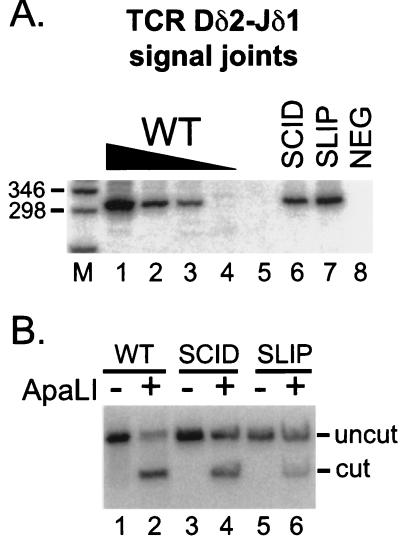
Figure 4.
Signal joint formation in SLIP mice. (A) TCR Dδ2-Jδ1 signal joints were amplified from thymocyte DNA preparations (the same DNA concentrations as in Fig. 3): wild-type titration (lanes 1–4), SCID (lane 6), and SLIP (lane 7). The expected size is 301 bp. A negative PCR control (all reagents without DNA) is shown in lane 8. Relevant sizes of the DNA marker (lane M) are indicated. (B) Signal joint PCR products were subjected to digestion with ApaLI. Wild type (lanes 1 and 2), SCID (lanes 3 and 4), and SLIP (lanes 5 and 6); undigested (lanes 1, 3, and 5), and ApaLI treated (lanes 2, 4, and 6). The expected sizes after digestion are 167 and 134 bp; only the larger product hybridizes to the probe.

