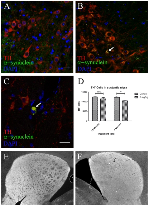
Figure 4. Alpha-synuclein accumulation and neuronal loss in the SNc after 3 but not 1.5 months intragastrical rotenone treatment.
(A–C, scale bars 20 µm; E–F, scale bars 200 µm). A, B, C, immunostaining against TH, alpha-synuclein and DAPI on SNc sections from 1.5 months control (A) and 3 months (B–C) treated mice. Arrow in B, alpha-synuclein small inclusions inside TH+ neurons. Arrow in C, large alpha-synuclein inclusion (|>8.14 µm) inside a dopamineric neuron in the SN. D, stereological quantification (n = 3) of TH+ neurons in the SN from control and treated mice. Asterisk, P<0.05. Number of neurons was determined based on the optical fractionator principle using StereoInvestigator software (MicroBrightField Inc., Williston, USA). Each column represents total number of TH+ neurons in the SN in 1.5 and 3 months control and treated mice. Graph shows mean ± s.e.m. E, F, TH immunostaining on striatum in 1.5 months control (E) and 3 months treated (F) mice.

