Fig. 2.
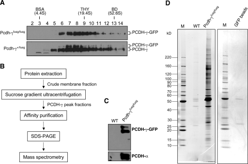
Affinity purification of PCDH-γ protein complexes. A, SG analysis of PCDH-γ-GFPs complexes from Pcdh-γ-GFPfusg/fusg and Pcdh-γ-GFP+/fusg mice (P0). PCDH-γ-GFPs and wild-type PCDH-γs are indicated. B, the flow chart of purification procedures. C, Western blot analysis of PCDH-γ-GFP immune complexes purified from Pcdh-γ-GFPfusg/fusg brains (P0–P5). An equal number of wild-type (WT) mouse brains served as a negative control for the purification. D, SYPRO Ruby-stained SDS-PAGE of the purified PCDH-γ-GFP immune complexes (left). To examine the possible protein contamination from anti-GFP-agarose beads, an equal amount of empty beads was eluted followed by SDS-PAGE and SYPRO Ruby staining (right). Only two faint bands, IgG heavy and light chains, were detected. M indicates the lanes loaded with marker proteins. THY, thyroglobulin; BD, blue dextran.

