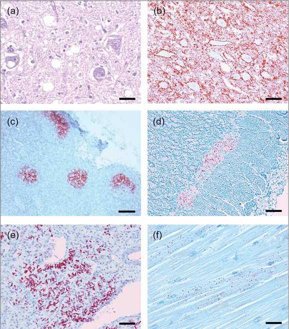
Figure 2.
Immunohistochemistry (IHC) detection of PrPCWD using MAb F99/97.6.1 in various tissues of orally inoculated red deer (Cervus elaphus elaphus) with CWD.
- Brain, medulla oblongata, obex, dorsal motor nucleus of vagus nucleus. There is moderate diffuse vacuolation of the neuropil and also occasional presence of vacuoles within neuronal perikarya. Hematoxylin & eosin. Bar = 30 μm.
- Brain, medulla oblongata, obex. There is extensive accumulation of PrPCWD (stained red) predominantly in the neuropil and with prominent perineuronal labelling. IHC. Bar = 70 μm.
- Tonsil. Detection of PrPCWD in tonsillar lymphoid follicles. IHC. Bar = 275 μm.
- Colon, myenteric plexus. IHC. Bar = 70 μm.
- Adrenal gland, medulla. IHC. Bar = 70 μm.
- Heart, ventricular septum. IHC. Bar = 40 μm.

