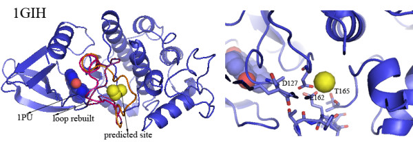
Figure 4.

The predicted calcium-binding site in the structure of human cell division protein kinase 2 (PDB ID: 1GIH) and the experimentally observed site in the structural homolog of 1GIH. 1GIH binds to 1PU (sphere colored by element, gray blue for carbon, dark blue for nitrogen, and red for oxygen). The structure of one large loop 149-164 (residues 149-164, sequence: ARAFGVPVRTYTHEVV) near the 1PU binding site is unstructured. Three possible loop conformations built by modeling methods are shown in red, pink and orange. The rebuilt loops locate at the domain interface and cover the binding pocket of 1PU. FEATURE identifies a calcium-binding site in the presence of the rebuilt loops. The close-up view shows the close associations between the predicted site and three residues F127, E162 and T165. The predicted site is 13 Å away from 1PU.
