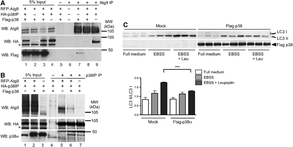
Figure 6.
Overexpression of p38α inhibits autophagy and competes with mAtg9 for binding to HA–p38IP. (A) HEK293A cells were transfected with RFP–mAtg9 and Flag–p38 (lanes 2 and 7), RFP–mAtg9, Flag–p38 and HA–p38IP (lanes 3 and 8), or RFP–mAtg9 and HA–p38IP (lanes 4 and 9). RFP–mAtg9 was immunoprecipitated with an anti-mAtg9 antibody, the co-immunoprecipitated HA–p38IP was detected with an anti-HA antibody, and co-immunoprecipitated Flag–p38 was detected with an anti-Flag antibody. RFP–mAtg9 was detected with an anti-Atg9 antibody. (B) HEK293A cells were transfected with RFP–Atg9 and HA–p38IP (lanes 1 and 5), RFP–mAtg9, Flag–p38, and HA–p38IP (lanes 2 and 6), or Flag–p38 and HA–p38IP (lanes 3 and 7). HA–p38IP was immunoprecipitated with an anti-HA antibody, the co-immunoprecipitated RFP–Atg9 was detected with an anti-Atg9 antibody, and co-immunoprecipitated Flag–p38 was detected with an anti-p38α antibody. HA–p38IP was detected with an anti-HA antibody. (C) HEK293A cells were transfected with Flag–p38α for 24 h. Cells were incubated in either full medium, EBSS, or EBSS plus leupeptin for 2 h, lysed, and analysed for endogenous LC3 lipidation using an anti-LC3 antibody, and immunoblotted with anti-Flag antibody. LC3 lipidation was quantified as the amount of LC3II/LC3I (data are represented as mean±s.e.m. of triplicates, n=2 experiments, EBSS control versus EBSS with Flag-p38α, ***P=0.0026).

