Figure 6.
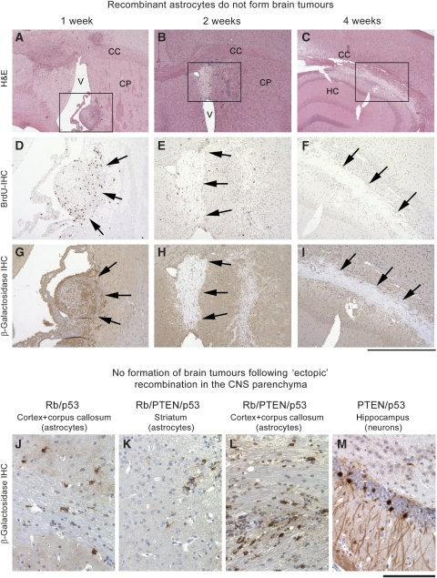
Recombinant astrocytes do not form brain tumours. In vitro recombined astrocytes (PTEN/Rb/p53) were implanted into the striatum of recipient mice from identical genetic background. At 1 week after grafting (A, D, G) there are several nodules of spindle shaped cells in several locations, such as the ventricle, attached to the striatum (boxed area), immunostaining for BrdU shows frequent proliferating cells. (G) Recombined cells show expression of β-galactosidase (arrows) in the graft, but only background staining of the neuropil. After 2 weeks, the grafts have largely degenerated into gliotic scar tissue and show only infrequent proliferating residual cells (E, BrdU IHC). (H) β-galactosidase IHC shows absence of staining in these degenerate grafts, notably, this tissue gives less background stain than CNS tissue. At 4 weeks (C, F, I) only scar tissue remains and no more proliferating cells are seen (F), and β-galactosidase immunoreactivity is absent. Scale bar: 1.3 mm (A–C), 550 μm (D–I). (J–M) Ectopic injection of Adeno-cre recombines grey and white matter astrocytes but does not cause their neoplastic transformation in vivo. Occasionally, hippocampal neurons were recombined, but no tumours formed.

