Figure 7.
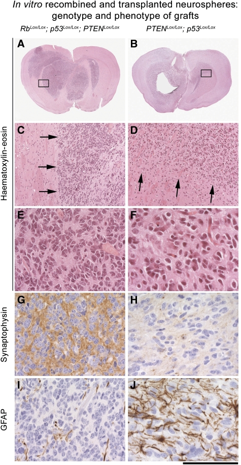
In vitro-recombined neurospheres generate tumours with a phenotype that resembles those generated by in vivo recombination. Left column (A, C, E, G, I): tumours derived from injection of Rb/p53/PTEN neurospheres. Right column (B, D, F, H, J): tumours derived from injection of p53/PTEN neurospheres. Panels (A–F) haematoxylin and eosin. Arrows in (C) and (D) show a well-demarcated (C) border in PNET or a diffuse infiltration into the CNS (D). Panels (G, H): synaptophysin is expressed in PNET (G) but not in gliomas (H). Panels (I, J) GFAP is not expressed in PNET (I) but clearly identifiable in neoplastic astrocytes in gliomas (J). Scale bar: 4 mm (A, B), 350 μm (C, D) and 90 μm (E–J).

