
Figure 1. Diagramatic location of 162 different mutations in the KCNH2 potassium channel involving 858 subjects.
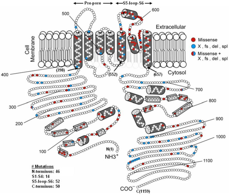
The α subunit involves the N-terminus (NH3+), six membrane-spanning segments, and the C-terminus portion (COO-). The numbers in the parentheses refer to the position of the amino acid beginning at the N-term position (1), the beginning of the transmembrane non-pore S1-S4 sequence (398), the beginning of the transmembrane S5-loop-S6 sequence (552), the end of the transmembrane S6 sequence (657), and at the C-term end position (1159). The open circles represent individual amino acids, the red circles indicate the missense mutations, and the blue circles indicate non-missense mutations. The cylinders represent putative α-helical segments, and the bars represent putative β-sheets.