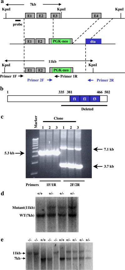
Fig. 1.
Targeted disruption of the KLF11 gene. (a) Schematic representations of the wild-type KLF11 locus, targeting vector, and the targeted allele. (b) Sequences that are deleted from the KLF11 gene. (c) PCR analysis of KLF11 targeted ES cells. The primer set 1F and 1R detects a 5.3-kb band for the targeted allele and no band for the wild-type allele. The primer set 2F and 2R detects a 3.7-kb band for the targeted allele and a 7.1-kb band for the wild-type allele. (d) Southern blot analysis of targeted ES cells. DNA isolated from ES cells was digested with KpnI. The wild-type allele is detected as a 7-kb fragment. The targeted allele is detected as an 11-kb fragment. (e) Southern blot analysis of mice generated from a KLF11+/− cross. DNA isolated from mice tail was digested with KpnI. The wild-type allele is detected as a 7-kb fragment. The targeted allele is detected as an 11-kb fragment.

