
Fig. 1.
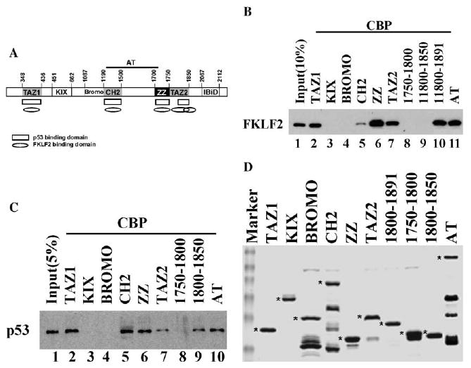
CBP interacts with FKLF2 and p53 through distinct domains. (A) The schematic representation of CBP domain structure. FKLF2 and p53 interacting domains of CBP are indicated as oval and rectangle. (B) GST pull-down assays were carried out by incubating 1 μl of whole cell extracts prepared from COS cells expressing Myc-tagged FKLF2 protein with 1 μg of purified GST–CBP fusion proteins immobilized on glutathione–agarose beads as indicated. The presence of FKLF2 was detected by immunoblotting using anti-Myc 9E10 monoclonal antibody and chemiluminescence. (C) GST pull-down assays were carried out by incubating 1 μl of whole cell extracts prepared from COS cells expressing Flag-tagged FKLF2 protein with 1 μg of purified GST–CBP fusion proteins immobilized on glutathione–agarose beads as indicated. The presence of p53 was detected by immunoblotting using anti-Flag M2 monoclonal antibody and chemiluminescence. (D) Coomassie blue staining of the GST–CBP fusion proteins was used in the assay. The full-length proteins are marked by an asterisk.