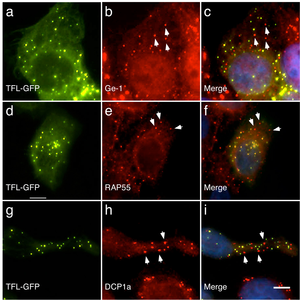
Figure 1.
p58TFL-GFP does not localize to P-bodies. When expressed in Hep-2 cells, p58TFL-GFP localized to dot-like structures throughout the cell (green, a, d, and g). However, p58TFL-GFP did not co-localize with antibodies directed against P-body markers Ge-1, RAP55, or DCP1a (red, b, e, and g). Merge of panels a and b, d and e, and g and h are shown in c, f and i, respectively. White arrows indicate representative P-bodies that lack p58TFL-GFP. DAPI staining in c, f, and i (blue) indicate the location of cell nuclei. White bar indicates 5 µm.

