
Figure 1. A cartoon representation of the peptide-based biosensor for the c-Abl kinase.
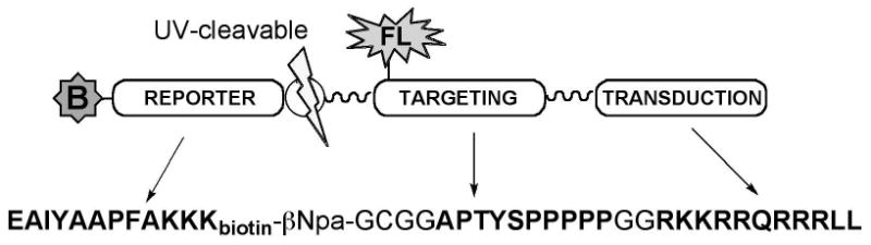
The substrate contains a biotinylated “reporter” module that is phosphorylated by c-Abl; a “targeting” module binds the Abl SH3 domain; and a “transduction” module (the TAT peptide), to aid the peptide in crossing the membrane barrier. The reporter and targeting sequences are connected via a photocleavable linker to allow release of the reporter from the backbone of the biosensor.