
Figure 2. Cellular uptake and distribution of substrate peptide.
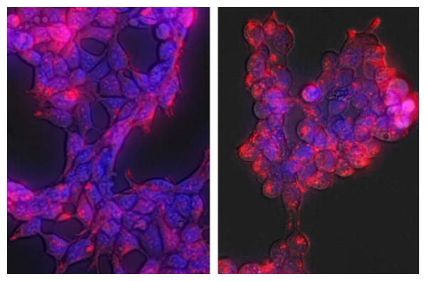
HEK293 cells were treated with Alexafluor-555-labeled Abl biosensor peptide (red, 25 μM) for 15 min (left panel) or 4 h (right panel) at 37 °C in media. Nuclei in the live cells were stained with Hoechst 33342 (blue). Cells were washed with PBS and visualized using a Zeiss Axiovert fluorescence microscope within 5 min. Peptide was rapidly taken up and distributed throughout the cell (15 min), persisting up to 4 h.