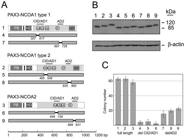
Figure 9.
Analyses of transforming activities of PAX3-NCOA1 and PAX3-NCOA2 fusion proteins and deletion mutants. A. Schematic structure of PAX3-NCOA1, PAX3-NCOA2 and deletion mutants. Solid bars with amino acid position displays the regions deleted. B. Immunoblot analyses of PAX3-NCOA1, PAX3-NCOA2 and deletion mutants in NIH3T3-Tet-19.1.E. C. Soft agar colony assay of pcDNA4/TO vector (1), pcDNA4/TO-PAX3-NCOA1 type 1, pcDNA4/TO-PAX3-NCOA1 type 2, and pcDNA4/TO-PAX3-NCOA2 transduced NIH3T3-Tet-19.1 cells. Cells were incubated in the presence of 1μg/ml tetracycline and scored after 14 days. Three plates were counted for each construct.

