
Figure 1.
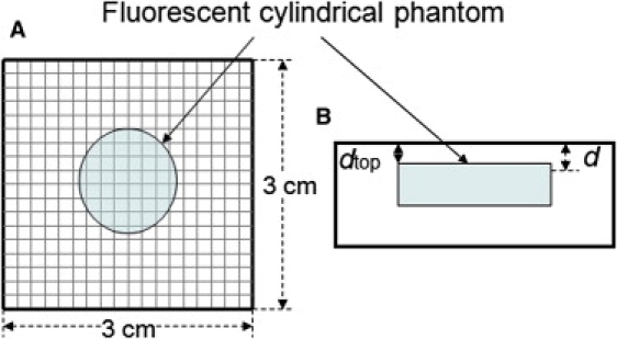
(A) Scanning geometry is composed of a source and a detector with 3-mm fixed separation and scans every 2 mm. The diameter and the thickness of this cylindrical phantom were 15 mm and 8 mm, respectively. The imaged area is 3 × 3 cm2. (B) Side view of Fig. 1 A.