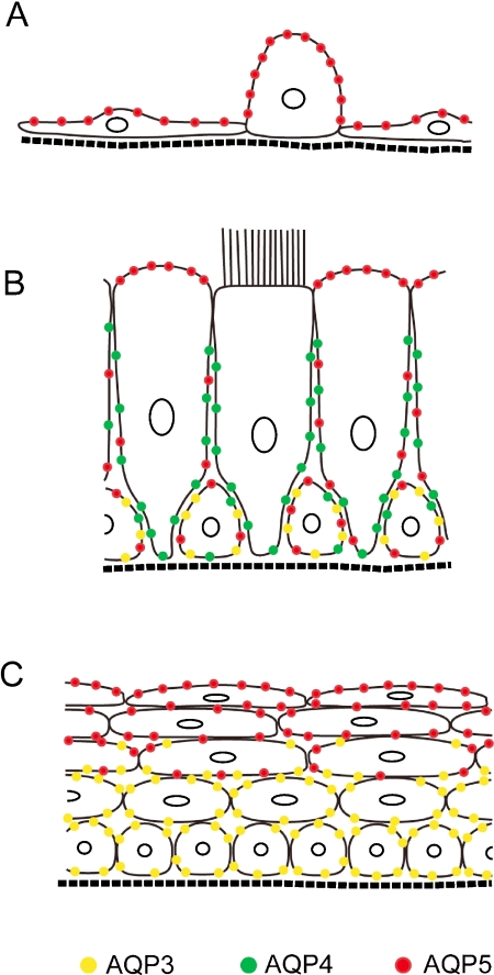
Fig. 7.
Schema summarizing the distribution patterns of AQP3, AQP4, and AQP5 in the mouse respiratory epithelia. A: Alveolar epithelium containing exquisitely thin type I and a cuboidal type II cells. B: Tracheal pseudostratified columnar epithelium composed of ciliated, non-ciliated, and basal cells. C: Stratified squamous epithelium in the upper airway.

