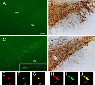
Fig. 2.
Comparison of FJC staining (A and C) and TH immunostaining (B and D) between the contralateral side (A and B) and ipsilateral side (C and D) in the SN at 3 days after intrastriatal 6-OHDA injection. High magnification view of the highlighted box in C (inset). Cell bodies of the degenerating neurons induced by 6-OHDA were double labeled with IF using TH (E and H) and FJC (F and I) in the SNc and were round (E–G) or shrunken (H–J). G and J represent mergers of E and F, and H and I respectively. SN, substantia nigra; SNc, substantia nigra pars compacta; SNr, substantia nigra pars reticularis. Bars=100 µm (A–D) and 10 µm (inset in C and E–J).

