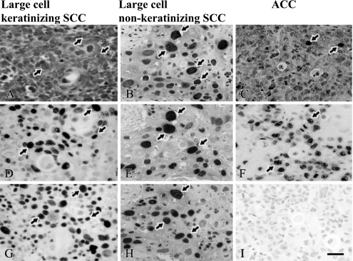
Fig. 4.
A and C: Localization of HPV 16; B: HPV cocktail; D–F: Ki-67 and G–I: p63 in cervical cancer. (A, D and G) The panels were obtained from large cell keratinizing SCC, (B, E and H) from large cell non-keratinizing SCC and (C, F and I) ACC of each adjacent sections. (D, G, E and H) Ki-67 and p63-positive cells were abundant in SCC; however, (I) p63 was negative in ACC. (A and D, B and E) HPV 16 and HPV cocktail were co-expressed with Ki-67 in SCC. Arrows indicate positive cells for HPV 16, Ki-67 and p63 in cervical cancer. Bar=20 µm.

