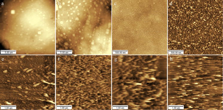
Fig. 3.
AFM height images of various biological samples determined with variety of scanner types, scanning modes and scanning forces on fresh cleaved mica. (a&b) K562 cell with 55μm and 125μm scanner respectively, (c&d) phosphatidylcholine monolayer (Langmuir film) with tapping and contact mode respectively, (e~h) phosphatidylcholine monolayer with contact mode in different contact force.

