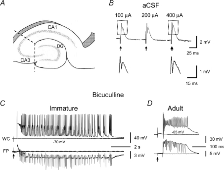
Figure 1. Prolonged population bursts after bicuculline treatment in CA3 minislices of immature versus adult rats.
A, diagram showing CA3 minislices used in this study. Dashed lines indicate knife cuts to isolate the CA3 area from CA1 and dentate gyrus. B shows that in normal aCSF, synaptic stimulation in CA3 minislices from immature rats only elicited a field EPSP and a single population spike and never evoked epileptiform bursts, regardless of the stimulus intensity (the three responses were evoked at 100 μA, 200 μA and 400 μA). Arrows indicate stimulations. Boxed parts in B are shown in expanded scales below. Note the difference in the amplitude of population spikes at different stimulus intensities (i.e. 100 μA and 400 μA). C, simultaneous whole-cell (WC, current-clamp mode) and field-potential (FP) recordings showing evoked population bursts in bicuculline (30 μm) in CA3 minislices from immature rats. These bursts typically contained an initial depolarization followed by rhythmic afterdischarges lasting for several seconds. D, by contrast, the evoked population bursts that occurred in CA3 slices of mature rats were composed of a single burst that lasted for a few hundred milliseconds.

