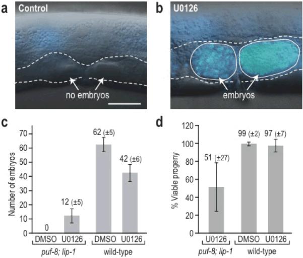
Figure 2. Small-molecule induced oocytes are functional.

(a–b) Whole animals were imaged by DIC and DAPI (blue) staining after treatment with control (DMSO) or U0126. (a) DMSO-treated mutant lacking embryos (dashed outline indicating empty uterus). (b) U0126-treated mutant with embryos containing numerous DAPI-stained nuclei. (c) Number of embryos produced by mutants and wild type treated with DMSO or U0126. (d) Percentage of embryos that developed to adulthood after parents were treated with DMSO of U0126. Approximately half of the embryos from chemically-induced oocytes developed to adulthood. Data are presented as mean +/− s.d. Scale bar, 10 μm.
