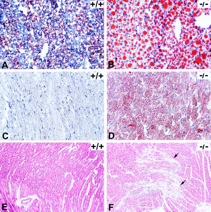
Figure 3.
Histopathology on LCAD +/+ (normal control) and LCAD −/− (deficient mice). (A) +/+ and (B) −/−: Oil-Red-O staining of frozen liver sections from 4- to 5-wk-old mice after an 18- to 20-hr fast. Abundant large lipid droplets present in the LCAD-deficient mice compared with minimal lipid accumulation in normal control. (C) +/+ and (D) −/−: Oil-Red-O staining of frozen hearts from 14- to 16-wk-old mice after an 18- to 20-hr fast. Note lipid accumulation in all cardiomyocytes of the LCAD-deficient mouse as compared with the LCAD-normal control. (E) +/+ and (F) −/−: Hematoxylin/eosin-stained heart sections from 16-wk-old, nonfasted male mice. Note prominent myocardial degeneration and fibrosis (arrows) present in the LCAD −/− mice (F). (A and B, ×25, C and D, ×40, and E and F, ×10.)

