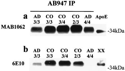
Figure 2.
Identification of the apoE–Aβ aggregate. (a) Human brain soluble fractions immunoprecipitated with the anti-apoE antibody AB947 show a broad band of 34–40 kDa in both AD and control (CO) subjects that is immunodetectable with the monoclonal antibody MAB1062, specific for apoE; the lower portion of this band comigrates with human plasma apoE. Note that the 34- to 40-kDa band is less prominent in AD than in control brains. (b) The same samples immunodetected with the antibody 6E10 (specific for Aβ residues 6–10) show an ≈40-kDa band, corresponding to the upper portion of the band detected with the antibody MAB1062, that comigrates with an apoE–Aβ complex formed in vitro (XX) (see Materials and Methods). The ≈40-kDa band is less prominent in all the AD brains, and in the apoE ɛ4 homozygous (4/4) subject it is visible only after a prolonged exposure (not shown).

