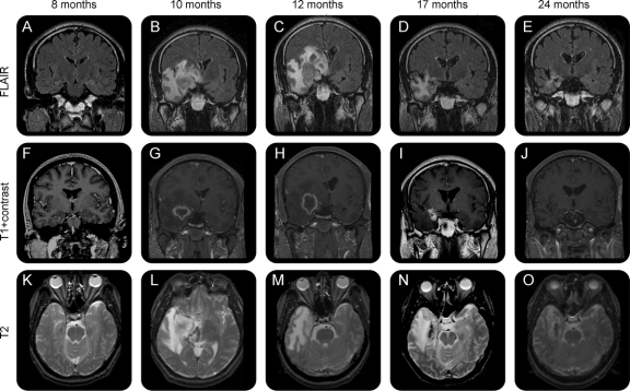
Figure 2 Development of radiologic changes in a representative patient treated with a 24-Gy dose
Fluid-attenuated inversion recovery coronal (A–E), T1 coronal with gadolinium (F–J), T2 axial (K–O). Abnormal T2 prolongation involving the right frontal and temporal lobes demonstrating central heterogeneity at the level of the right medial temporal lobe. Severe vasogenic edema resulting in a mild increase in midline shift. The central area of heterogeneity has peripheral enhancement. By 12 months, the swelling of the temporal lobe resulted in almost 1 centimeter of midline shift on the coronal view. The T2 abnormality is largely limited to the white matter. The corresponding volume of abnormal T2 hyperintensity is 703 mL, the volume of contrast enhancement is 31.4 mL, the T2 hyperintensity anatomic extent score is 5 (extends to parietal and frontal lobes), and the mass effect score is 3 (local swelling with narrowing of the ambient cistern). At 24 months, there was significant reduction in the extent of T2 hyperintensity (102 mL, score 1), with residual T2 prolongation only in the right anterior parahippocampal gyrus, fusiform gyrus, and extending superiorly along the right temporal stem into the inferior subinsular cortex. There was interval decrease in mass effect at 24 months (mass effect score = 0). The right temporal lobe now appears atrophic compared to the left temporal lobe.

