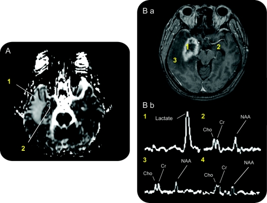
Figure 4 Potential mechanisms of radiosurgery for medial temporal lobe epilepsy revealed by magnetic resonance diffusion and spectroscopy
(A) Diffusion magnetic resonance at 12 months after treatment shows increased diffusion throughout the temporal lobe (1). There is also an area of decreased diffusion, and heterogeneity in the medial temporal lobe (2) suggesting ongoing delayed ischemic changes. (B) Proton magnetic resonance spectroscopy. T1-weighted axial MRI after gadolinium administration for spectroscopy voxel measurements (B.a). Spectra obtained for individual voxels (1 mL) selected in the following areas (B.b): (1) radiosurgery target region in mesial temporal lobe at 12 months, (2) contralateral mesial temporal lobe at 12 months, (3) peri-target region edema at 12 months, and (4) target region in mesial temporal lobe before radiosurgery treatment (not shown, but same position as (1) before treatment). The right medial temporal lobe demonstrates a prominent lactate peak in the area of contrast enhancement, and the near absence of the N-acetylaspartate signal compared to other regions or before treatment.

