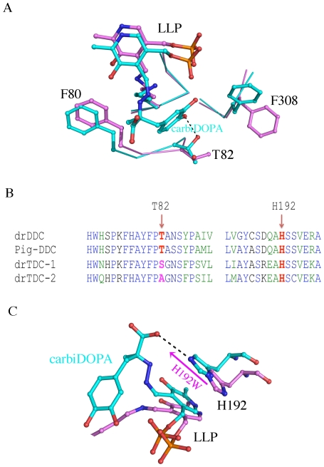
Figure 6. The roles of T82 and H192.
A, Superposition of drDDC structure onto pig DDC structure shows that T82 of drDDC is a putative substrate binding residue. Residues from pig DDC are colored in cyan and those from drDDC are colored in magenta. B, Alignment of drDDC, pig DDC, Drosophila tyrosine decarboxylase-1 (drTDC-1) and Drosophila tyrosine decarboxylase-2 (drTDC-2). The corresponding residues of T82 and H192 are labeled. C, Superposition of drDDC structure onto pig DDC structure shows the H192 of drDDC is a putative substrate and cofactor binding residue. Residues from pig DDC are colored in cyan and those from drDDC are colored in magenta.

