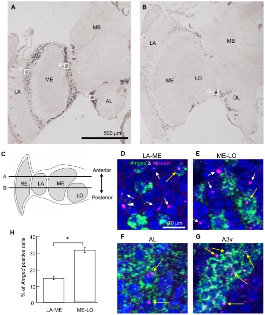
Figure 1. Amgad and kakusei expression in the worker brain.
A, B. Expression of Amgad was detected by in situ hybridization using the rostral (A) and caudal (B) worker brain sections. Left hemispheres of coronal sections are shown. Note the strong Amgad signals in the optic lobe and antennal lobe neurons. C. Schematic drawing of the optic lobe of the worker brain and the position of the rostral (A) and caudal (B) sections. Dorsal view. Anterior is top. D–G. Double fluorescent in situ hybridization of kakusei (magenta) and Amgad (green) in the seizure (sz)-induced worker brain. The nuclei stained with DAPI are shown in blue. White arrows indicate kakusei (+) and Amgad (−) nuclei, and yellow arrows indicate kakusei (+) and Amgad (+) nuclei. Sometimes, nuclei with two intranuclear foci for kakusei were observed (white arrow head). The positions of (D)–(G) are indicated by the white squares in (A) and (B). H. The proportion of Amgad (+) cells was different between LA-ME and ME-LO. *: P<0.0001, Welch's t-test. Abbreviations: AL, antennal lobe; DL, dorsal lobe; LA, lamina; LO, lobula; ME, medulla; MB, mushroom body; RE, retina.

