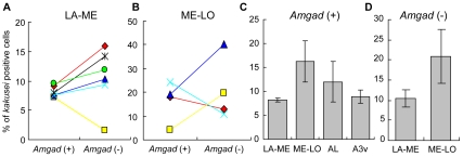
Figure 2. Percentage of kakusei-positive Amgad (+) and (−) cells in the seizure (sz)-induced bees.
The data from each bee is shown by the same symbol and connected by a line. A, B. Percentage of kakusei-positive cells in LA-ME (A) and ME-LO (B). There was no significant difference in the kakusei expression between Amgad (+) and (−) cells in either region. C, D. Comparison of the percentages of kakusei-positive Amgad (+) (C) and (−) cells in various brain regions (D). No significant difference in the kakusei expression was detected among these brain regions.

