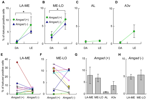
Figure 5. kakusei expression in the brains of dark-adapted (DA) and light-exposed (LE) bees.
A–D. Changes in kakusei expression induced by the light-exposure. kakusei expression was significantly increased in LA-ME (A) and ME-LO (B) (*: P<0.01, respectively). The increase was similar between Amgad (+) and (−) cells. No significant increase in AL (C) or A3v (D) was detected. The percentages of kakusei-positive Amgad (+) and (−) cells did not differ significantly in LA-ME (E) or ME-LO (F). G, H. The percentage of kakusei-positive cells was not significantly different among various brain regions.

