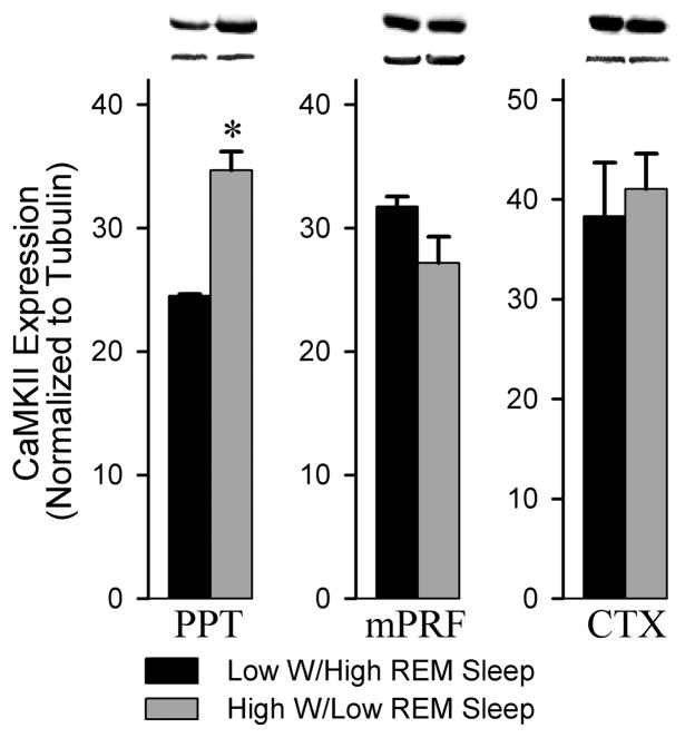
Figure 2.
Analysis of CaMKII expression in the PPT, mPRF and cortex of low W/high REM sleep and high W/low REM sleep subjects. Western blot analysis of CaMKII expression in the PPT revealed a significant 29.2% reduction in the low W/high REM sleep group compared to the high W/low REM sleep condition (t = 18.48, p = 0.003). In contrast, western blot analysis of CaMKII expression in the adjacent mPRF (t = 1.874, n.s.) and the cortex (t = 0.4347, n.s.) revealed no difference between the low W/high REM sleep and high W/low REM sleep conditions. All analyses of CaMKII expression are normalized against α-tubulin. * indicates a significant difference <0.01.

