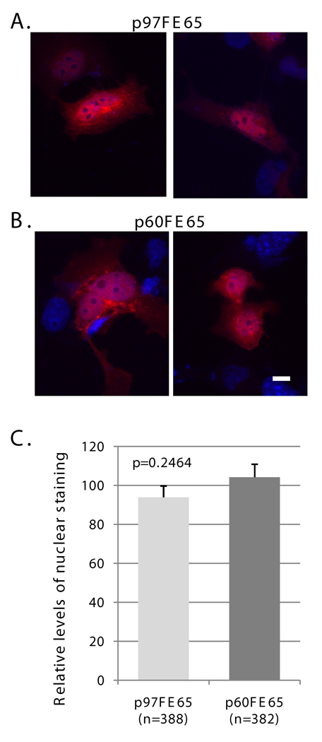
Fig. 3. p60FE65 is able to translocate to the nucleus.
A–B: COS cells were transfected with pcDNA3.1-p97FE65 (A) or pcDNA3.1-KOFE65(M327L) (B) and stained with antibody FE518 (red). Nuclei were visualized with DAPI (blue). C: Relative levels of nuclear p97FE65 (A) and p60FE65 (B) were determined by fluorescence quantitation of stained nuclei in p97FE65-positive cells (n =388), and p60FE65 [KOFE65(M327L)]-positive cells (n=382), respectively. Data were presented as mean ± SE, and analyzed by two-tailed t-test. The results showed that there was no significant difference in FE65 levels in the nuclei of the two groups of cells. Size bar equals 10 microns.

