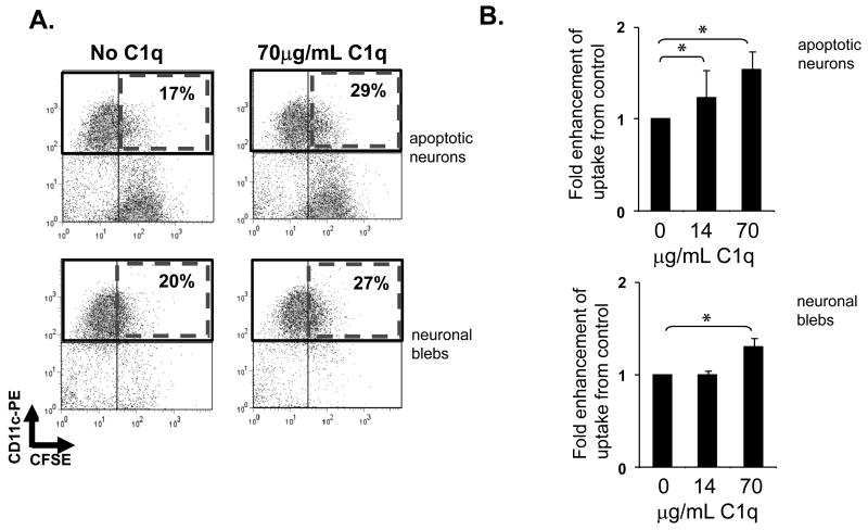
Figure 3. Microglial uptake of apoptotic neurons and neuronal blebs.
CFSE-labeled apoptotic neurons and neuronal blebs were added to rat microglia in the presence or absence of C1q as described in Materials and Methods. Uptake of CSFE-labeled targets by anti CD11b-labeled microglia was assessed by flow cytometry, with a single representative experiment depicted in (A) and expressed as average fold enhancement of uptake over control levels in the absence of C1q (B). +/- SD. * p<0.05 ANOVA, n=5 (apoptotic neurons), n=4 (neuronal blebs)

