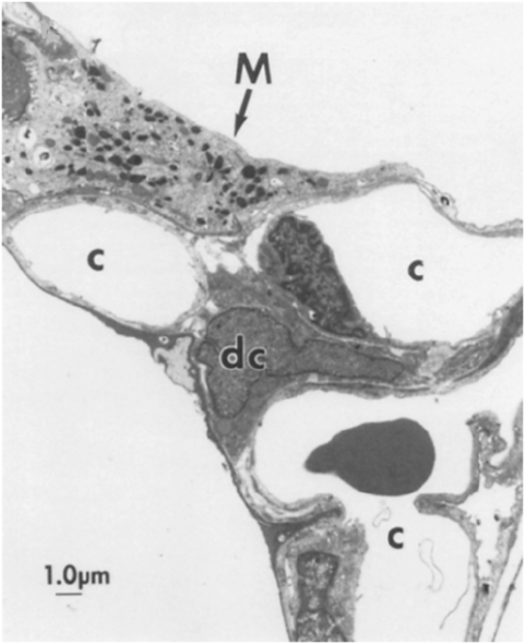
Figure 1.
Interstitial dendritic cells (DCs) and alveolar macrophages in the alveolus. Electron micrograph of rat alveolar septal junction showing a large macrophage (M) containing many electron-dense vacuoles, spread upon the type I epithelial lining of an alveolus, and a smaller DC (dc) with an irregularly shaped indented nucleus, located in the interstitial space. c, capillary. Reprinted with permission from Reference 45.

