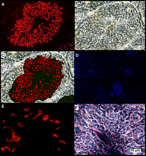
FIG. 2.
Zonadhesin distribution and expression in mature stallion testis. Zonadhesin was localized by immunofluorescence on sections of testis from a 3-yr-old stallion using antisera to pig zonadhesin holoprotein. A) Zonadhesin immunoreactivity in a seminiferous tubule (epifluorescence). Note the strong labeling in the region of developing spermatids. B) Phase contrast view of the tubule shown in A. C) Panels A and B merged. D) Epifluorescence view of a seminiferous tubule stained with fluorescent secondary antibody only (no zonadhesin antiserum). E) Higher magnification view of zonadhesin immunoreactivity in elongating spermatids near the lumen of a tubule (epifluorescence). F) Phase contrast view of the field shown in E. Bars indicate relative magnification of the images.

